Dissection of muscles of male perineum and pelvic diaphragm
Superior fascia of urogenital diaphragm
Stanford holds the copyright to the David L. Bassett anatomical images and has assigned
Creative Commons license Attribution-Share
Alike 4.0 International to all of the images.
For additional information regarding use and permissions,
please contact the Medical History Center.
Image #174-5
Dissection of muscles of male perineum and pelvic diaphragm
Superior fascia of urogenital diaphragm
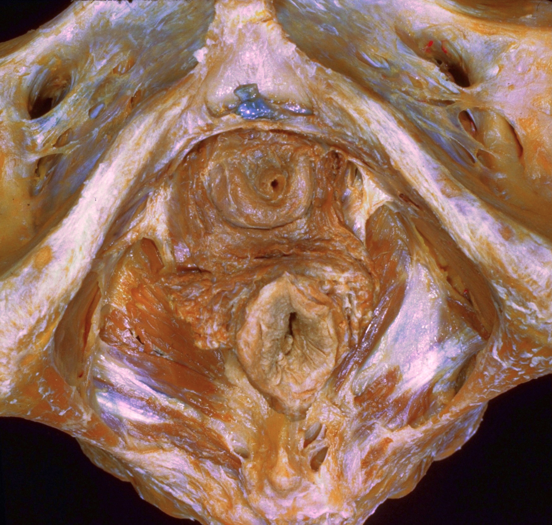
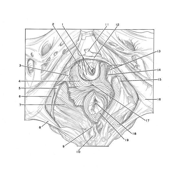
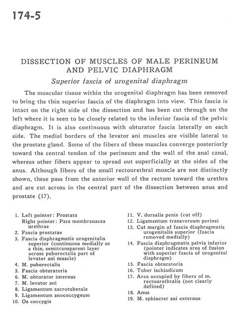
The muscular tissue within the urogenital diaphragm has been removed to bring the thin superior fascia of the diaphragm into view. This fascia is intact on the right side of the dissection and has been cut through on the left where it is seen to be closely related to the inferior fascia of the pelvic diaphragm. It is also continuous with obturator fascia laterally on each side. The medial borders of the levator ani muscles are visible lateral to the prostate gland. Some of the fibers of these muscles converge posteriorly toward the central tendon of the perineum and the wall of the anal canal, whereas other fibers appear to spread out superficially at the sides of the anus. Although fibers of the small rectourethral muscle are not distinctly shown, these pass from the anterior wall of the rectum toward the urethra and are cut across in the central part of the dissection between anus and prostate (17).
- Left pointer: Prostate Right pointer: Membranous part of urethra
- Prostatic fascia
- Superior fascia of urogenital diaphragm (continuous medially as a thin, semitransparent layer across puborectalis part of levator ani muscle)
- Puborectalis muscle
- Obturator fascia
- Obturator internus muscle
- Levator ani muscle
- Sacrotuberous ligament
- Anococcygeal ligament
- Coccyx
- Dorsal vein of the penis (cut off)
- Transverse perineal ligament
- Cut margin of superior fascia of urogenital diaphragm (fascia removed medially)
- Inferior fascia of pelvic diaphragm (pointer indicates area of fusion with superior fascia of urogenital diaphragm)
- Obturator fascia
- Ischial tuberosity
- Area occupied by fibers of rectourethralis muscle (not clearly defined)
- Anus
- External anal sphincter muscle