Dissection of anal triangle and ischiorectal fossae
Close-up view of anus and nerves, vessels and fascia of ischiorectal fossae
Stanford holds the copyright to the David L. Bassett anatomical images and has assigned
Creative Commons license Attribution-Share
Alike 4.0 International to all of the images.
For additional information regarding use and permissions,
please contact the Medical History Center.
Image #173-5
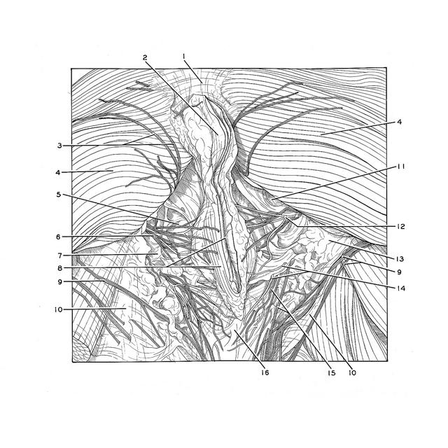
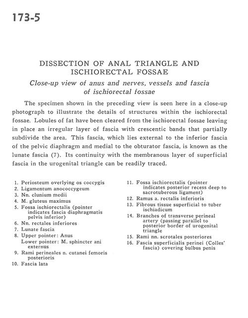
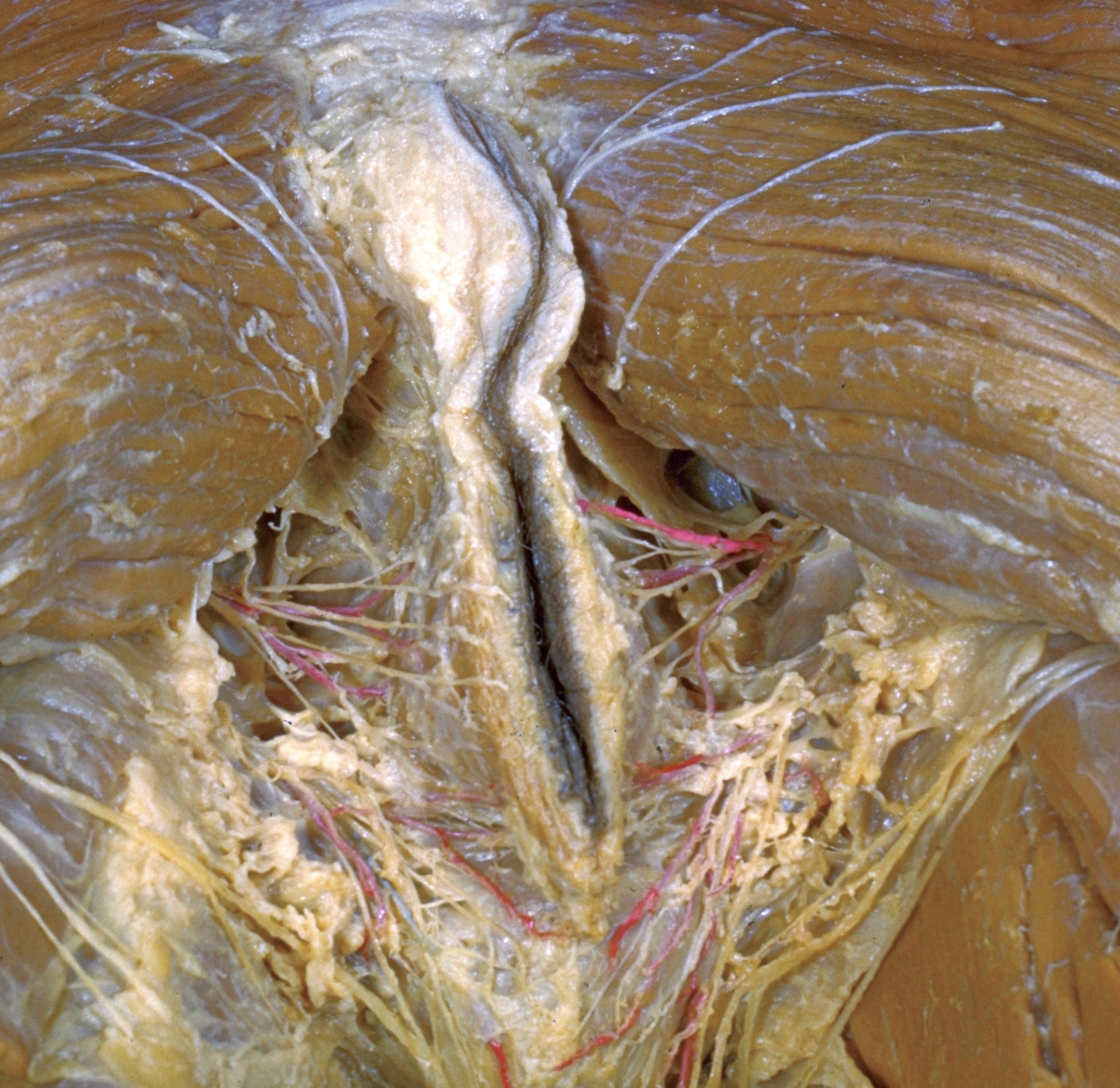
Dissection of anal triangle and ischiorectal fossae
Close-up view of anus and nerves, vessels and fascia of ischiorectal fossae
The specimen shown in the preceding view is seen here in a close-up photograph to illustrate the details of structures within the ischiorectal fossae. Lobules of fat have been cleared from the ischiorectal fossae leaving in place an irregular layer of fascia with crescentic bands that partially subdivide the area. This fascia, which lies external to the inferior fascia of the pelvic diaphragm and medial to the obturator fascia, is known as the lunate fascia (7). Its continuity with the membranous layer of superficial fascia in the urogenital triangle can be readily traced.
- Periosteum overlying coccyx
- Anococcygeal ligament
- Middle cluneal nerves
- Gluteus maximus muscle
- Ischiorectal fossa (pointer indicates inferior fascia of pelvic diaphragm)
- Inferior rectal nerves
- Lunate fascia
- Upper pointer: Anus Lower pointer: External anal sphincter muscle
- posterior perineal branches femoral cutaneous nerve
- Fascia lata
- Ischiorectal fossa (pointer indicates posterior recess deep to sacrotuberous ligament)
- Branch of inferior rectal artery
- Fibrous tissue superficial to tuber ischiadicum
- Branches of transverse perineal artery (passing parallel to posterior border of urogenital triangle)
- Branches of posterior scrotal nerves
- Supeficial perineal fascia (Colles' fascia) covering bulb of penis