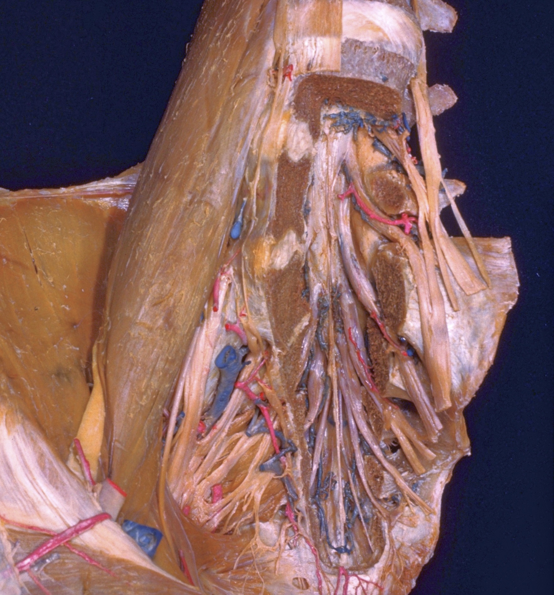
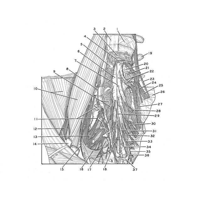
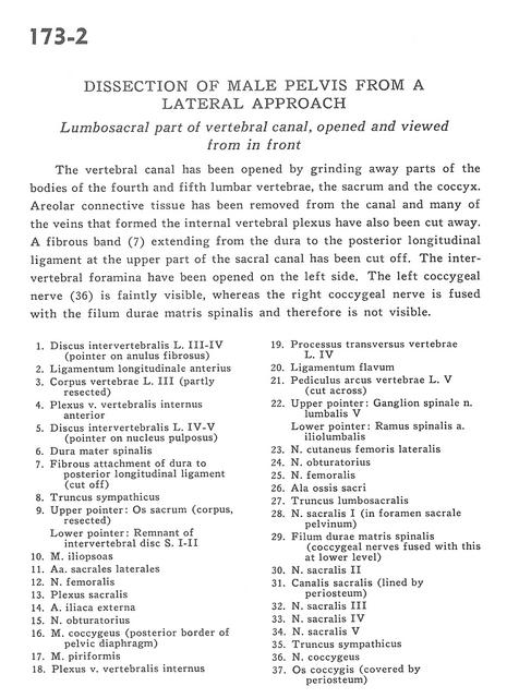
Dissection of male pelvis from a lateral approach
Lumbosacral part of vertebral canal, opened and viewed from in front
Stanford holds the copyright to the David L. Bassett anatomical images and has assigned
Creative Commons license Attribution-Share
Alike 4.0 International to all of the images.
For additional information regarding use and permissions,
please contact the Medical History Center.
Image #173-2
Dissection of male pelvis from a lateral approach
Lumbosacral part of vertebral canal, opened and viewed from in front
The vertebral canal has been opened by grinding away parts of the bodies of the fourth and fifth lumbar vertebrae, the sacrum and the coccyx. Areolar connective tissue has been removed from the canal and many of the veins that formed the internal vertebral plexus have also been cut away. A fibrous band (7) extending from the dura to the posterior longitudinal ligament at the upper part of the sacral canal has been cut off. The intervertebral foramina have been opened on the left side. The left coccygeal nerve (36) is faintly visible, whereas the right coccygeal nerve is fused with the filum durae matris spinalis and therefore is not visible.
- Intervertebral disc L. III-IV (pointer on anulus fibrosus)
- Anterior longitudinal ligament
- Body of vertebra L. III (partly resected)
- Anterior internal vertebral venous plexus
- Intervertebral disc L. IV-V (pointer on nucleus pulposus)
- Spinal dura mater
- Fibrous attachment of dura to posterior longitudinal ligament (cut off)
- Sympathetic trunk
- Upper pointer: Sacrum (corpus, resected) Lower pointer: Remnant of intervertebral disc S. I-lI
- Iliopsoas muscle
- Lateral sacral arteries
- Femoral nerve
- Sacral plexus
- External iliac artery
- Obturator nerve
- Coccygeus muscle (posterior border of pelvic diaphragm)
- Piriform muscle
- Internal vertebral venous plexus
- Transverse process vertebrae L. IV
- Ligamentum flavum
- Pedicle of arch of vertebra L. V (cut across)
- Upper pointer: Spinal ganglion lumbar nerve V Lower pointer: Spinal branch iliolumbar artery
- Lateral femoral cutaneous nerve
- Obturator nerve
- Femoral nerve
- Ala of sacrum
- Lumbosacral trunk
- Sacral nerve I (in anterior (pelvic) sacral foramen)
- Dural filum spinal cord (coccygeal nerves fused with this at lower level)
- Sacral nerve II
- Sacral canal (lined by periosteum)
- Sacral nerve III
- Sacral nerve IV
- Sacral nerve V
- Sympathetic trunk
- Coccygeal nerve
- Coccyx (covered by periosteum)