Dissection of male pelvis from a lateral approach
Pelvic plexus of right side, medial view
Stanford holds the copyright to the David L. Bassett anatomical images and has assigned
Creative Commons license Attribution-Share
Alike 4.0 International to all of the images.
For additional information regarding use and permissions,
please contact the Medical History Center.
Image #172-2
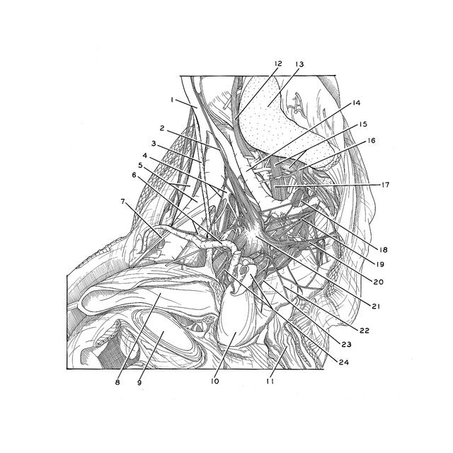
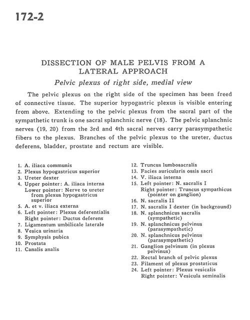
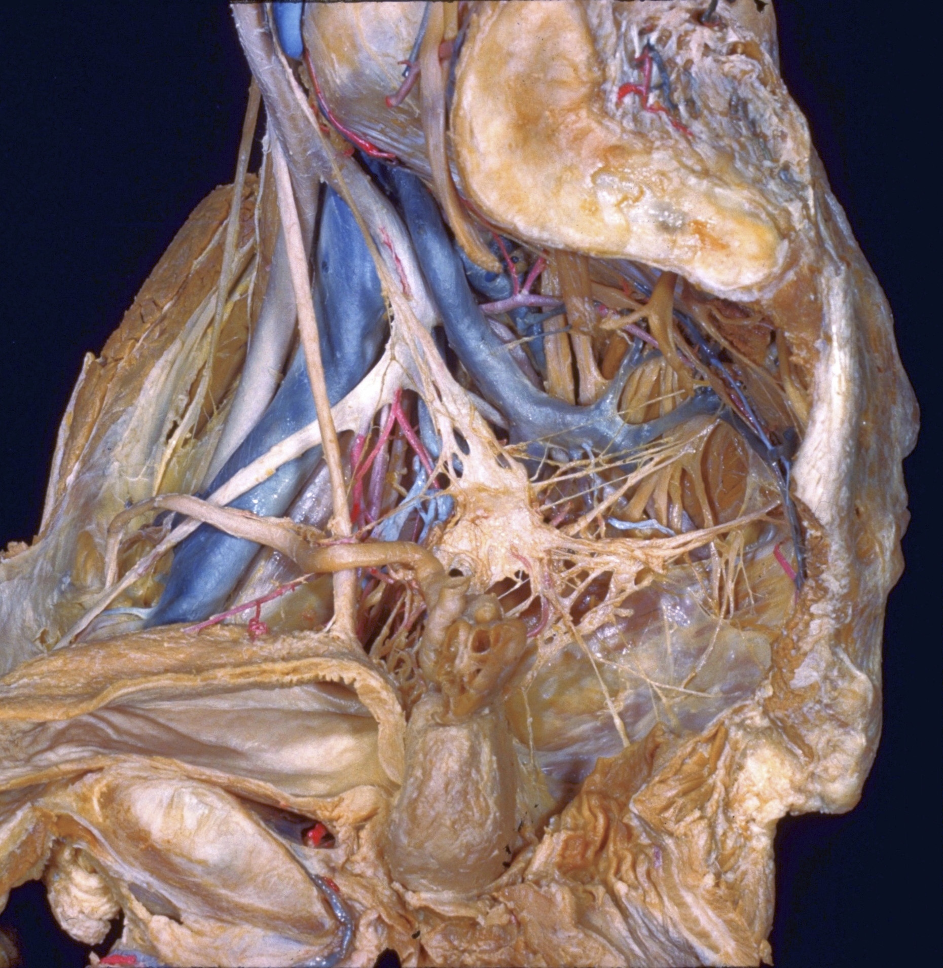
Dissection of male pelvis from a lateral approach
Pelvic plexus of right side, medial view
The pelvic plexus on the right side of the specimen has been freed of connective tissue. The superior hypogastric plexus is visible entering from above. Extending to the pelvic plexus from the sacral part of the sympathetic trunk is one sacral splanchnic nerve (18). The pelvic splanchnic nerves (19, 20) from the 3rd and 4th sacral nerves carry parasympathetic fibers to the plexus. Branches of the pelvic plexus to the ureter, ductus deferens, bladder, prostate and rectum are visible.
- Common iliac artery
- Superior hypogastric plexus
- Ureter right
- Upper pointer: Internal iliac artery Lower pointer: Nerve to ureter from superior hypogastric plexus
- External iliac artery and vein
- Left pointer: Deferential plexus Right pointer: Ductus deferens
- Lateral umbilical ligament
- Urinary bladder
- Pubic symphysis
- Prostate
- Anal canal
- Lumbosacral trunk
- Articular surface of sacrum
- Internal iliac vein
- Left pointer: Sacral nerve I Right pointer: Sympathetic trunk (pointer on ganglion)
- Sacral nerve II
- Sacral nerve I right (in background)
- Sacral splanchnic nerve (sympathetic)
- Pelvic splanchnic nerve (parasympathetic)
- Pelvic splanchnic nerve (parasympathetic)
- Pelvic ganglion (in pelvic plexus)
- Rectal branch of pelvic plexus
- Filament of prostatic plexus
- Left pointer: Vesical plexus Right pointer: Seminal vesicle