Dissection of male pelvis from a lateral approach
Anal canal opened, close-up anterolateral view
Stanford holds the copyright to the David L. Bassett anatomical images and has assigned
Creative Commons license Attribution-Share
Alike 4.0 International to all of the images.
For additional information regarding use and permissions,
please contact the Medical History Center.
Image #171-7
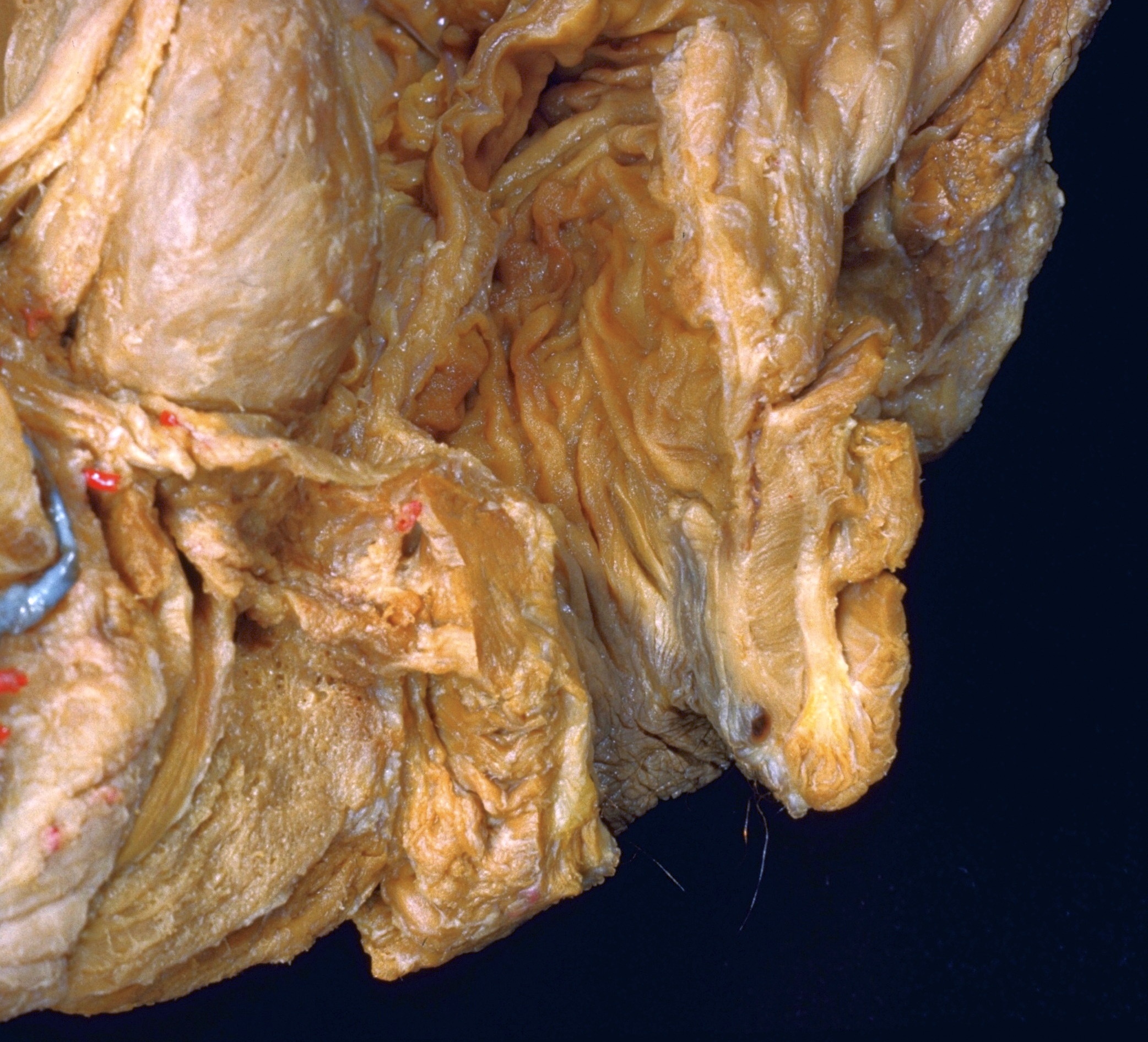
Dissection of male pelvis from a lateral approach
Anal canal opened, close-up anterolateral view
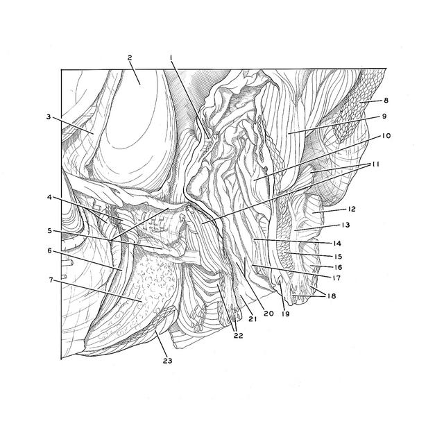
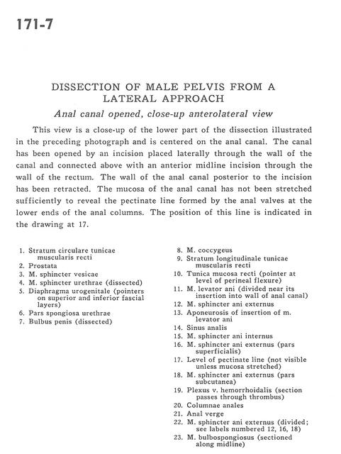
This view is a close-up of the lower part of the dissection illustrated in the preceding photograph and is centered on the anal canal. The canal has been opened by an incision placed laterally through the wall of the canal and connected above with an anterior midline incision through the wall of the rectum. The wall of the anal canal posterior to the incision has been retracted. The mucosa of the anal canal has not been stretched sufficiently to reveal the pectinate line formed by the anal valves at the lower ends of the anal columns. The position of this line is indicated in the drawing at 17.
- Circular layer of muscular tunic of rectum
- Prostate
- Vesical sphincter muscle
- Sphincter muscle of urethra (dissected)
- Urogenital diaphragm (pointers on superior and inferior fascial layers)
- Spongy part of urethra
- Bulb of penis (dissected)
- Coccygeus muscle
- Longitudinal layer of muscular tunic of rectum
- Mucosal tunic of rectum (pointer at level of perineal flexure)
- Levator ani muscle (divided near its insertion into wall of anal canal)
- External anal sphincter muscle
- Aponeurosis of insertion of Levator ani muscle
- Anal sinus
- Internal anal sphincter muscle
- External anal sphincter muscle (superficial part)
- Level of pectinate line (not visible unless mucosa stretched)
- External anal sphincter muscle (subcutaneous part)
- Hemorrhoidal venous plexus (section passes through thrombus)
- Anal columns
- Anal verge
- External anal sphincter muscle (divided see labels numbered 12, 16, 18)
- Bulbospongiosus muscle (sectioned along midline)