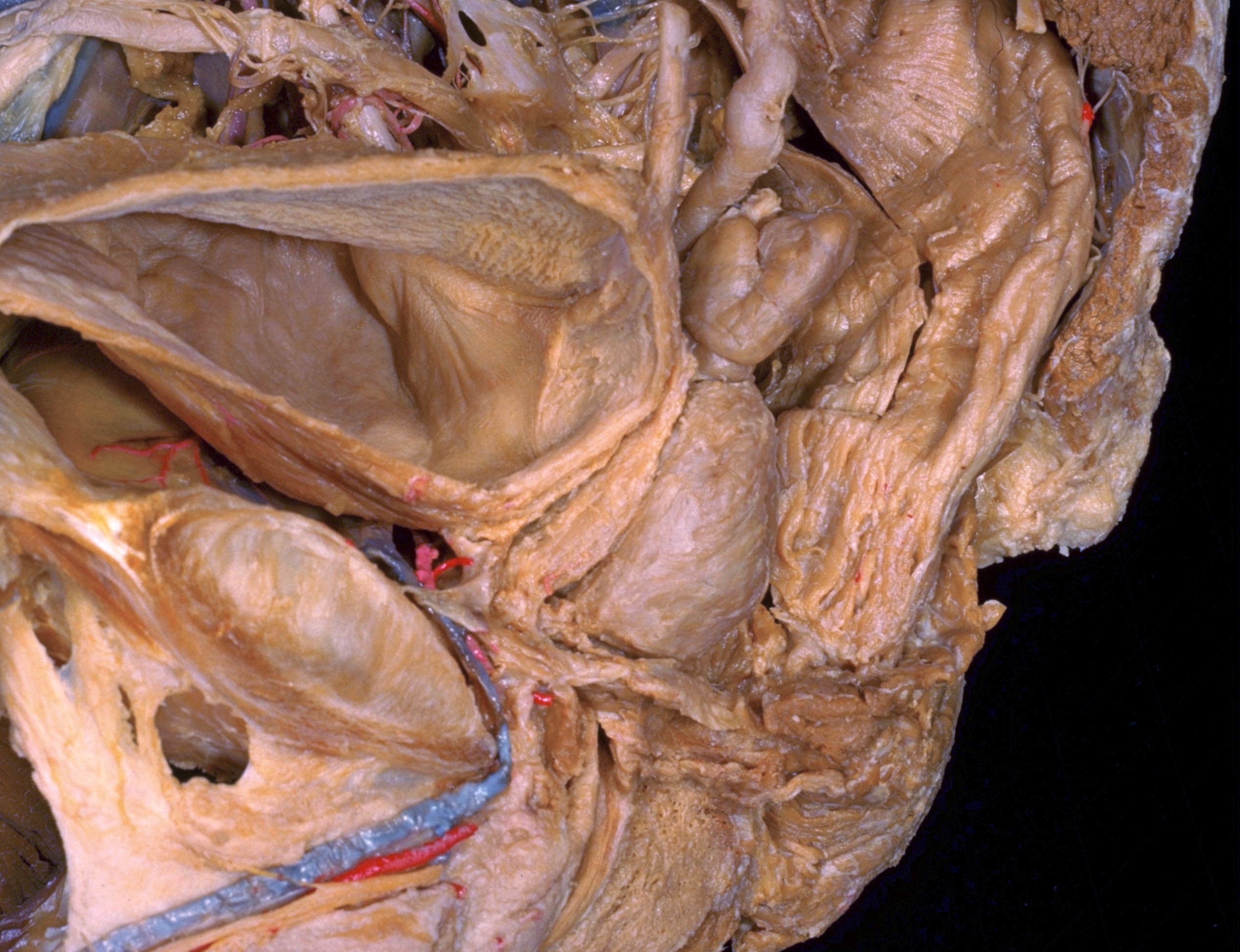
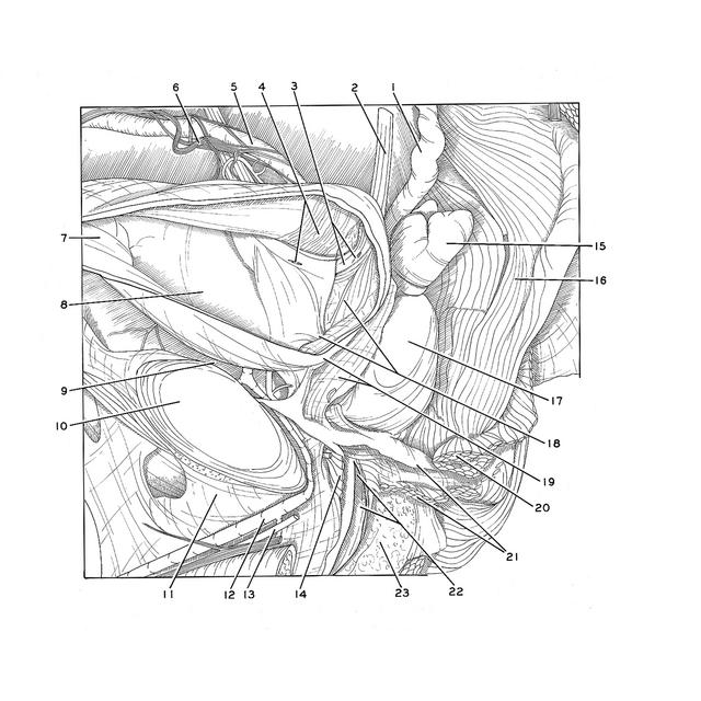
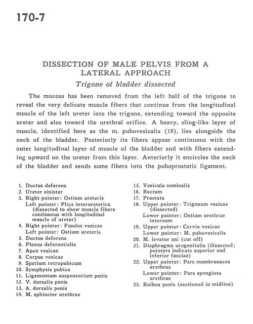
Dissection of male pelvis from a lateral approach
Trigone of bladder dissected
Stanford holds the copyright to the David L. Bassett anatomical images and has assigned
Creative Commons license Attribution-Share
Alike 4.0 International to all of the images.
For additional information regarding use and permissions,
please contact the Medical History Center.
Image #170-7
Dissection of male pelvis from a lateral approach
Trigone of bladder dissected
The mucosa has been removed from the left half of the trigone to reveal the very delicate muscle fibers that continue from the longitudinal muscle of the left ureter into the trigone, extending toward the opposite ureter and also toward the urethral orifice. A heavy, sling-like layer of muscle, identified here as the m. pubovesicalis (19), lies alongside the neck of the bladder. Posteriorly its fibers appear continuous with the outer longitudinal layer of muscle of the bladder and with fibers extending upward on the ureter from this layer. Anteriorly it encircles the neck of the bladder and sends some fibers into the puboprostatic ligament.
- Ductus deferens
- Ureter left
- Right pointer: Uterine opening Left pointer: Interureteric fold (dissected to show muscle fibers continuous with longitudinal muscle of ureter)
- Right pointer: Fundus of bladder Left pointer: Uterine opening
- Ductus deferens
- Deferential plexus
- Apex of bladder
- Body of bladder
- Retropubic space
- Pubic symphysis
- Suspensory ligament of the penis
- Dorsal vein of the penis
- Dorsal artery of penis
- Sphincter muscle of urethra
- Seminal vesicle
- Rectum
- Prostate
- Upper pointer: Trigone of urinary bladder (dissected) Lower pointer: Internal urethral opening
- Upper pointer: Cervix of bladder Lower pointer: Pubovesicalis muscle
- Levator ani muscle (cut off)
- Urogenital diaphragm (dissected; pointers indicate superior and inferior fasciae)
- Upper pointer: Membranous part of urethra Lower pointer: Spongy part of urethra
- Bulb of penis (sectioned in midline)