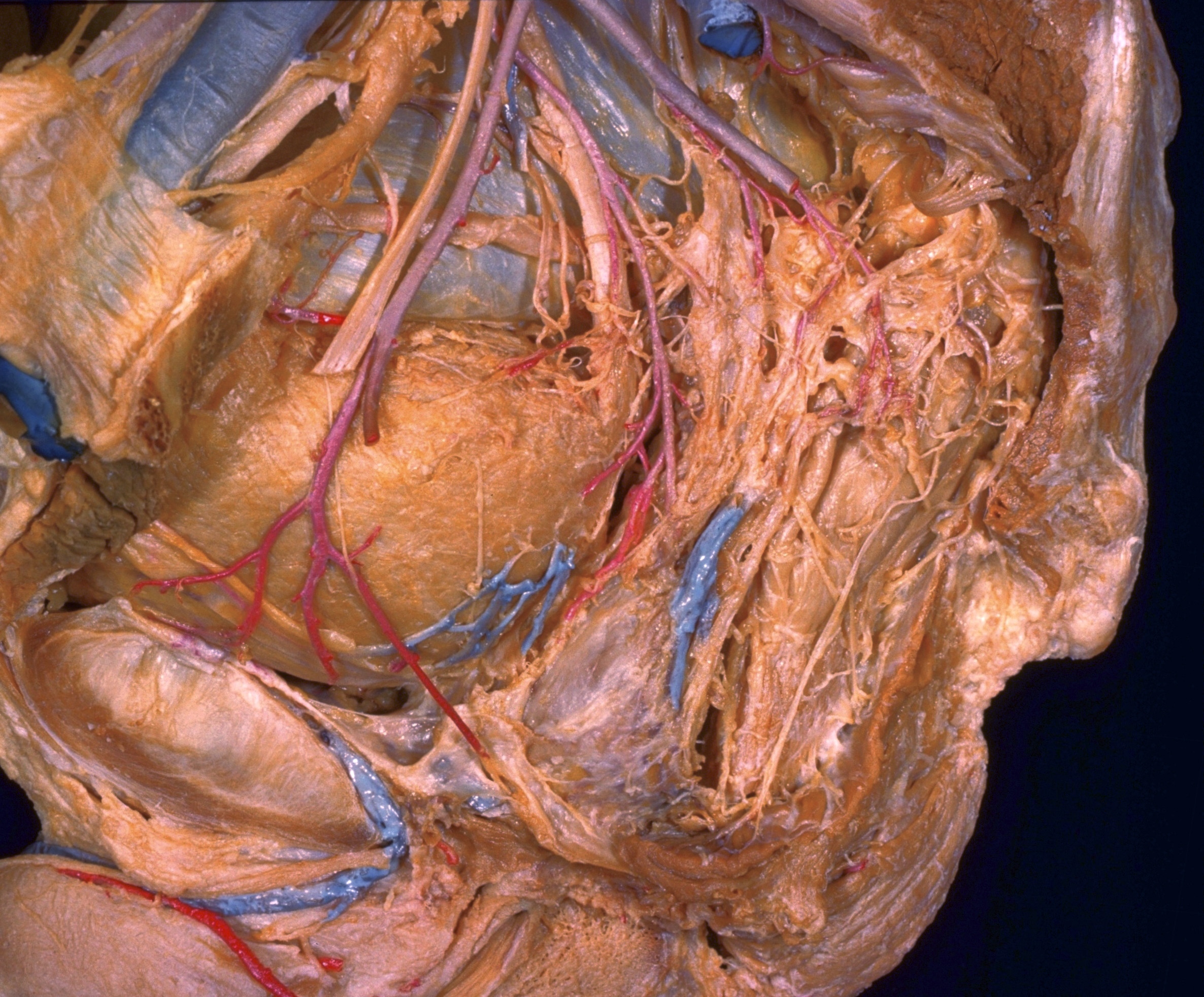
Dissection of male pelvis from a lateral approach
Pelvic organs in situ, close-up lateral view
Stanford holds the copyright to the David L. Bassett anatomical images and has assigned
Creative Commons license Attribution-Share
Alike 4.0 International to all of the images.
For additional information regarding use and permissions,
please contact the Medical History Center.
Image #169-7
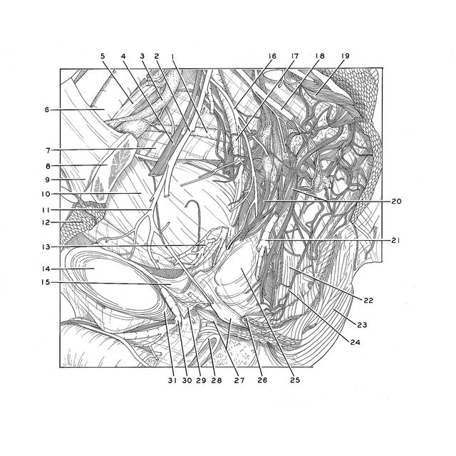
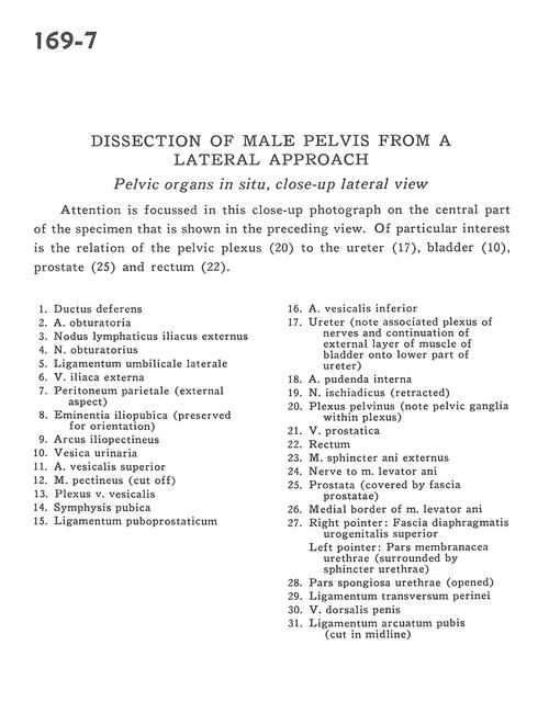
Dissection of male pelvis from a lateral approach
Pelvic organs in situ, close-up lateral view
Attention is focused in this close-up photograph on the central part of the specimen that is shown in the preceding view. Of particular interest is the relation of the pelvic plexus (20) to the ureter (17), bladder (10), prostate (25) and rectum (22).
- Ductus deferens
- Obturator artery
- External iliac lymph node
- Obturator nerve
- Lateral umbilical ligament
- External iliac vein
- Parietal peritoneum (external aspect)
- Iliopubic eminence (preserved for orientation)
- Iliopectineal arch
- Urinary bladder
- Superior vesical artery
- Pectineus muscle (cut off)
- Vesical venous plexus
- Pubic symphysis
- Puboprostatic ligament
- Inferior vesical artery
- Ureter (note associated plexus of nerves and continuation of external layer of muscle of bladder onto lower part of ureter)
- Internal pudendal artery
- Sciatic nerve (retracted)
- Pelvic plexus (note pelvic ganglia within plexus)
- Prostatic vein
- Rectum
- External anal sphincter muscle
- Nerve to Levator ani muscle
- Prostate (covered by prostatic fascia)
- Medial border of Levator ani muscle
- Right pointer: Superior fascia of urogenital diaphragm Left pointer: Membranous part of urethra (surrounded by sphincter of urethra)
- Spongy part of urethra (opened)
- Transverse perineal ligament
- Dorsal vein of the penis
- Pubic arcuate ligament (cut in midline)