Dissection of male pelvis from a lateral approach
General view of pelvic contents with hip bone removed
Stanford holds the copyright to the David L. Bassett anatomical images and has assigned
Creative Commons license Attribution-Share
Alike 4.0 International to all of the images.
For additional information regarding use and permissions,
please contact the Medical History Center.
Image #169-6
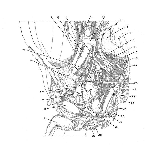
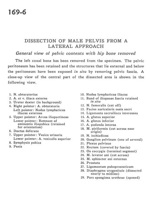
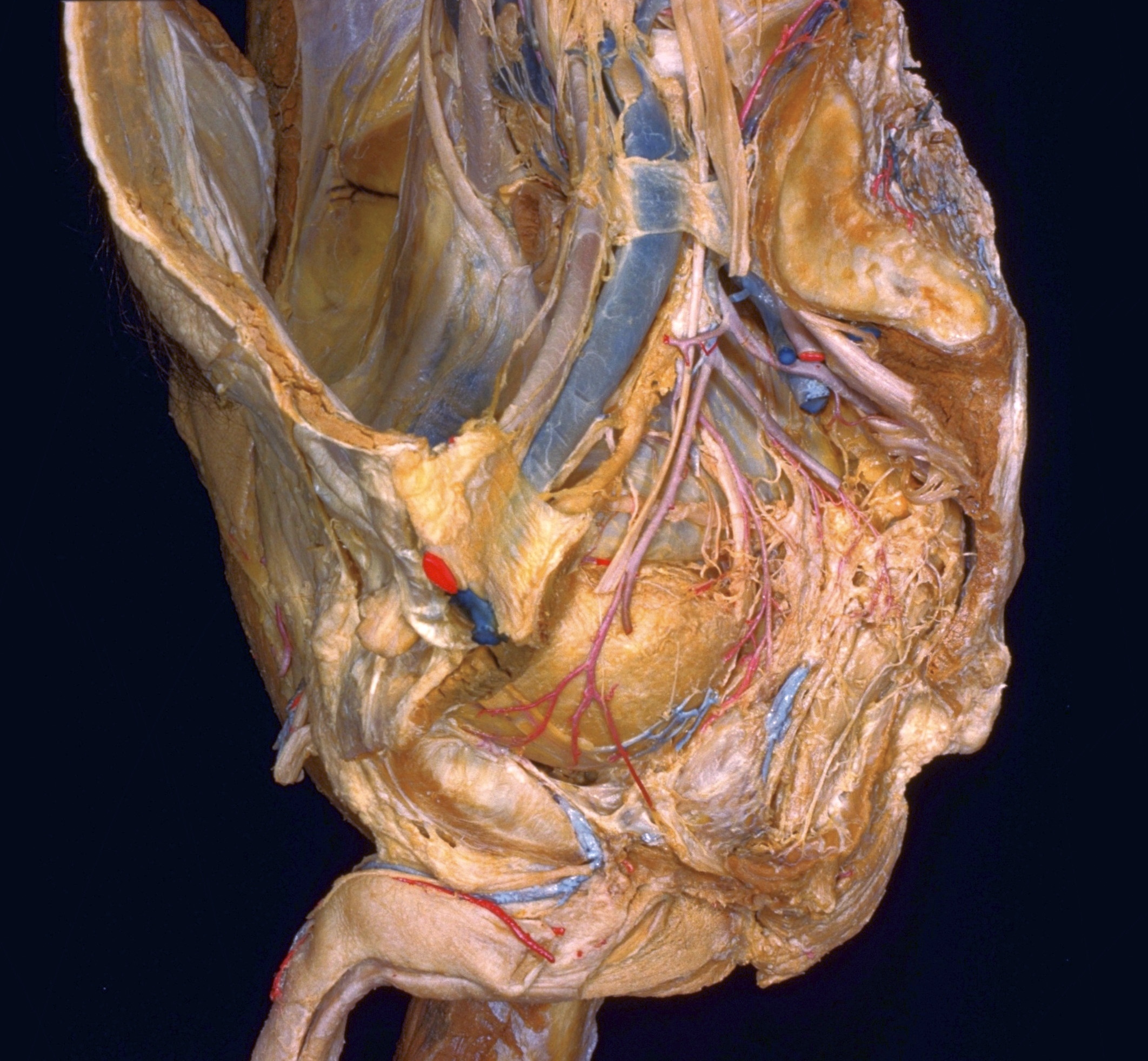
Dissection of male pelvis from a lateral approach
General view of pelvic contents with hip bone removed
The left coxal bone has been removed from the specimen. The pelvic peritoneum has been retained and the structures that lie external and below the peritoneum have been exposed in situ by removing pelvic fascia. A close-up view of the central part of the dissected area is shown in the following view.
- Obturator nerve
- External iliac artery and vein
- Ureter right (in background)
- Right pointer: Obturator artery Left pointer: External iliac lymph node
- Upper pointer: Iliopectineal arch Lower pointer: Remnant of iliopubic eminence (retained for orientation)
- Ductus deferens
- Upper pointer: Urinary bladder Lower pointer: Superior vesical artery
- Pubic symphysis
- Glans penis
- Iliac lymph node
- Band of iliopsoas fascia retained In situ
- Femoral nerve (cut off)
- Articular surface of sacrum
- Interosseous sacroiliac ligament
- Superior gluteal artery
- Inferior gluteal artery
- Internal pudendal artery
- Piriform muscle (cut across near origin)
- Sciatic nerve
- Pelvic ganglion (one of several)
- Pelvic plexus
- Rectum (covered by fascia)
- Coccyx (terminal segment)
- Levator ani muscle (cut across)
- External anal sphincter muscle
- Prostate
- Puboprostatic ligament
- Urogenital diaphragm (dissected nearly to midline)
- Spongy part of urethra (opened)