Male external genitalia and perineum
Membranous layer of superficial fascia in urogenital triangle
Stanford holds the copyright to the David L. Bassett anatomical images and has assigned
Creative Commons license Attribution-Share
Alike 4.0 International to all of the images.
For additional information regarding use and permissions,
please contact the Medical History Center.
Image #166-2
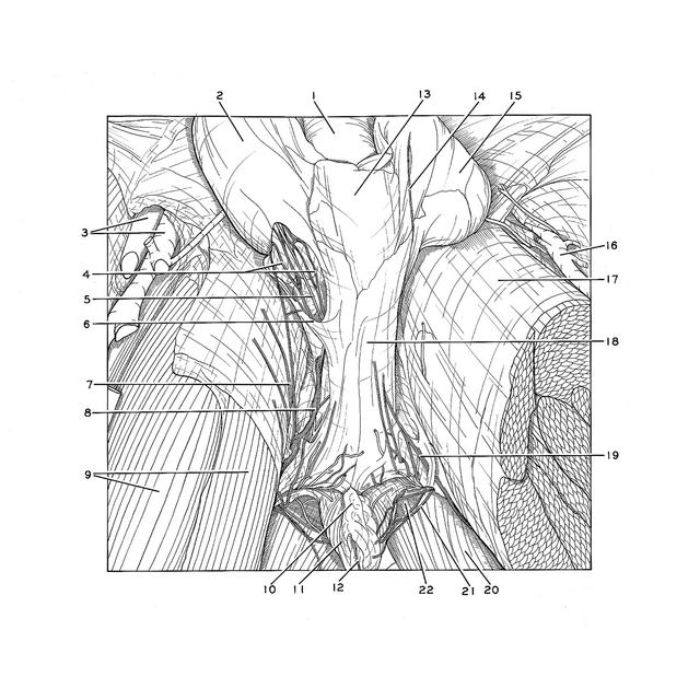
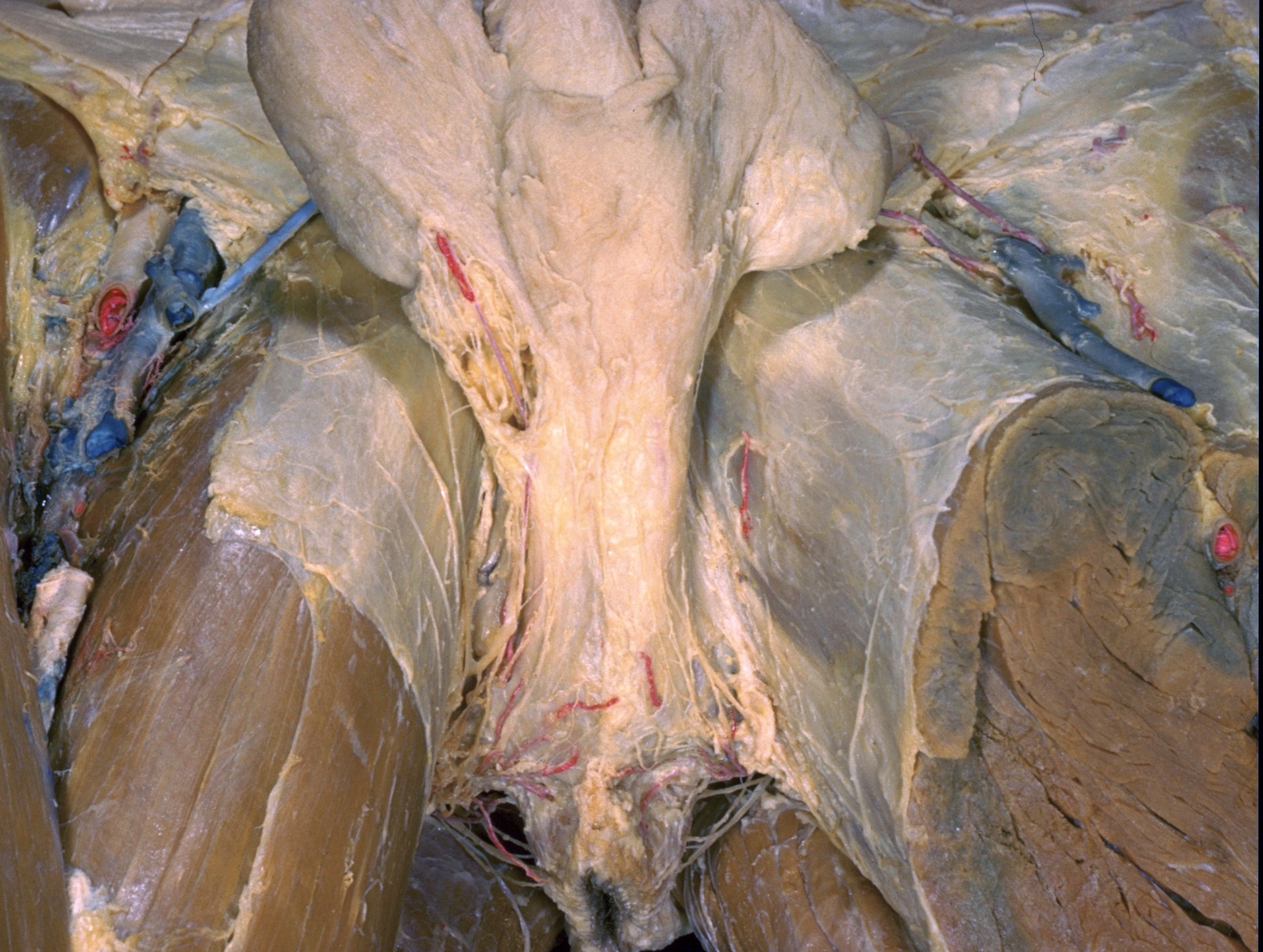
Male external genitalia and perineum
Membranous layer of superficial fascia in urogenital triangle
The specimen shown in view 165-2 is further explored in this preparation and in subsequent dissections of this series. The subcutaneous fat has been removed from the ischiorectal fossa. On the right side of the specimen an opening (6) has been made in the membranous layer of superficial fascia to expose some of the branches of the posterior scrotal artery and nerve. The scrotum has been retracted upward.
- Body of penis
- Dartos fascia (covering right testis)
- Femoral artery and vein
- Posterior scrotal nerves
- Posterior scrotal branch internal pudendal artery
- Margin of window cut through membranous layer of superficial fascia
- Perineal branch posterior femoral cutaneous nerve
- Perineal branch posterior femoral cutaneous nerve
- Upper pointer: Gracilis muscle Lower pointer: Adductor longus muscle
- Central tendon of perineum
- External anal sphincter muscle
- Anus
- Dartos fascia covering scrotal septum (spread out in retracting testicles)
- Terminal filament of posterior scrotal nerve
- Position of left testis (retracted with scrotum)
- Great saphenous vein
- Fascia lata
- Superficial perineal fascia (membranous layer)
- Perineal branch posterior femoral cutaneous nerve
- Gluteus maximus muscle
- Inferior rectal nerve
- Ischiorectal fossa