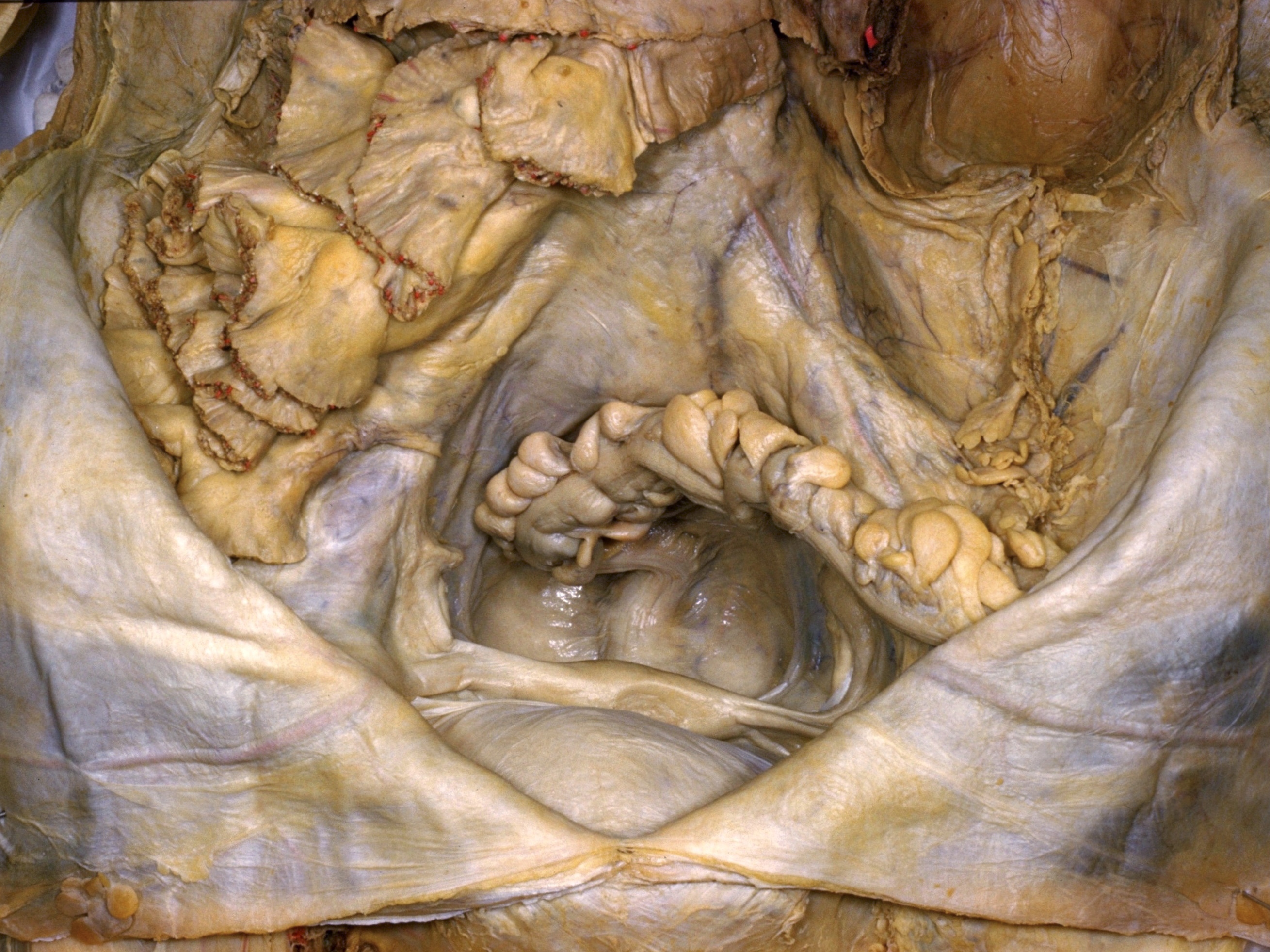
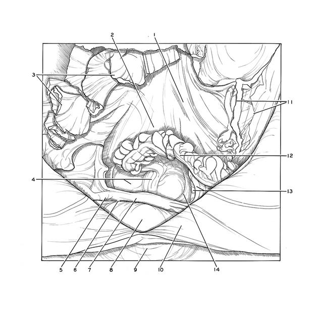
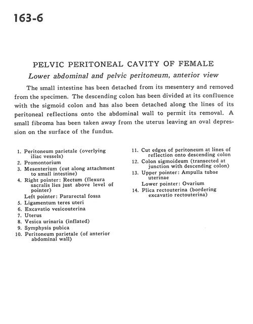
Pelvic peritoneal cavity of female
Lower abdominal and pelvic peritoneum, anterior view
Stanford holds the copyright to the David L. Bassett anatomical images and has assigned
Creative Commons license Attribution-Share
Alike 4.0 International to all of the images.
For additional information regarding use and permissions,
please contact the Medical History Center.
Image #163-6
Pelvic peritoneal cavity of female
Lower abdominal and pelvic peritoneum, anterior view
The small intestine has been detached from its mesentery and removed from the specimen. The descending colon has been divided at its confluence with the sigmoid colon and has also been detached along the lines of its peritoneal reflections onto the abdominal wall to permit its removal. A small fibroma has been taken away from the uterus leaving an oval depression on the surface of the fundus.
- Parietal peritoneum (overlying iliac vessels)
- Promontory
- Mesentery (cut along attachment to small intestine)
- Right pointer: Rectum (sacral flexure lies just above level of pointer) Left pointer: Pararectal fossa
- Ligamentum teres (of uterus)
- Uterovesical pouch
- Uterus
- Urinary bladder (inflated)
- Pubic symphysis
- Parietal peritoneum (of anterior abdominal wall)
- Cut edges of peritoneum at lines of reflection onto descending colon
- Sigmoid colon (transected at junction with descending colon)
- Upper pointer: Ampulla of uterine tube Lower pointer: Ovary
- Rectouterine fold (bordering rectouterine space)