Dissection of female pelvis from a lateral approach
Blood vessels and nerves of uterus, vagina and neck of bladder, anterior view
Stanford holds the copyright to the David L. Bassett anatomical images and has assigned
Creative Commons license Attribution-Share
Alike 4.0 International to all of the images.
For additional information regarding use and permissions,
please contact the Medical History Center.
Image #163-3
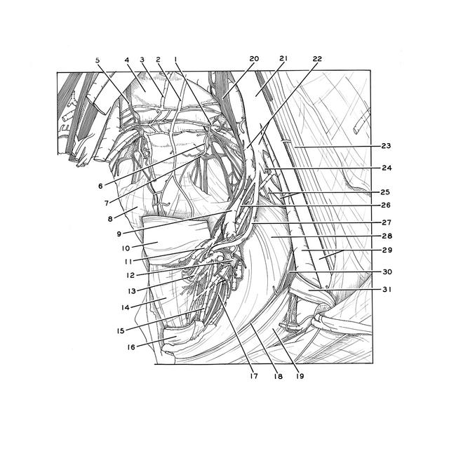

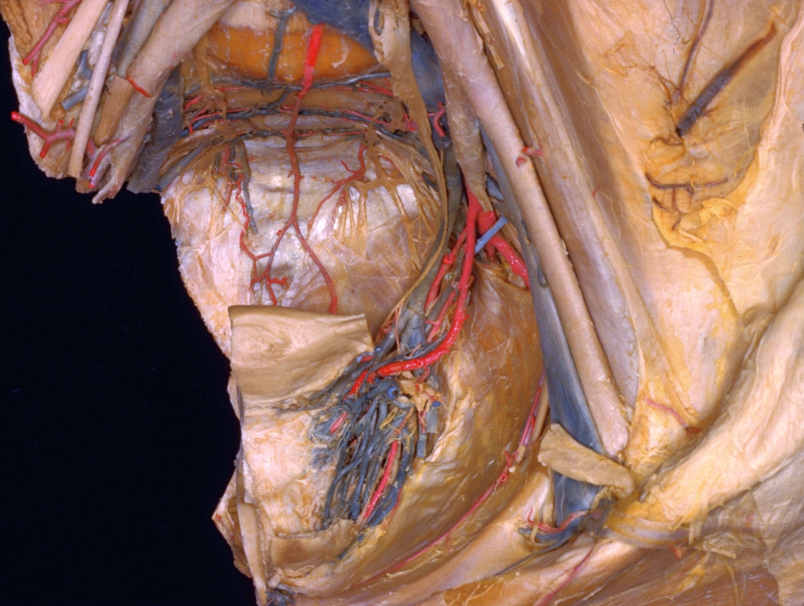
Dissection of female pelvis from a lateral approach
Blood vessels and nerves of uterus, vagina and neck of bladder, anterior view
The bladder has been transected close to the internal urethral opening and has been removed, together with the ureter, from the dissection. The anterior surfaces of the uterus and vagina are exposed to view.
- Left sympathetic trunk
- Middle sacral artery
- Middle sacral vein
- Intervertebral disc L. V - S. I
- Right sympathetic trunk
- Lateral sacral artery
- Sacral nerve IV
- Pelvic diaphragm (right half, cut close to sacral and coccygeal attachment)
- Uterine vein
- Uterus
- Uterine venous plexus
- Pelvic ganglion
- Vaginal venous plexus
- Vagina
- Vesical venous plexus
- Cervix of urinary bladder
- Inferior vesical artery
- Pubic branch of obturator artery
- Pectineal ligament
- Superior hypogastric plexus
- Common iliac artery
- Internal iliac artery and vein
- Psoas major muscle
- Lateral umbilical ligament
- Left pointer: Internal pudendal artery Right pointer: Inferior gluteal artery
- Vaginal artery
- Uterine artery
- Pelvic diaphragm
- External iliac artery and vein
- Obturator nerve
- Ligamentum teres (of uterus)