Dissection of female pelvis from a lateral approach
Interior of left side of pelvic cavity.
Stanford holds the copyright to the David L. Bassett anatomical images and has assigned
Creative Commons license Attribution-Share
Alike 4.0 International to all of the images.
For additional information regarding use and permissions,
please contact the Medical History Center.
Image #162-4
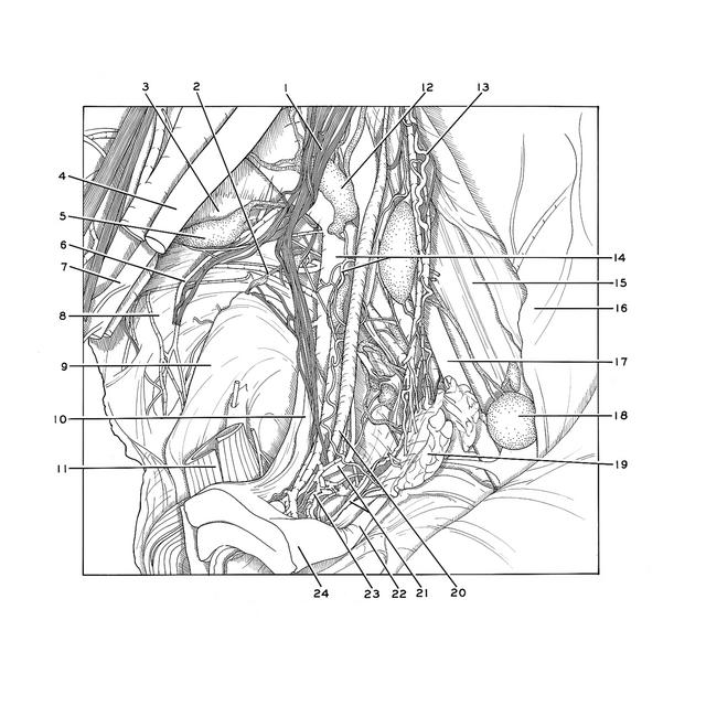
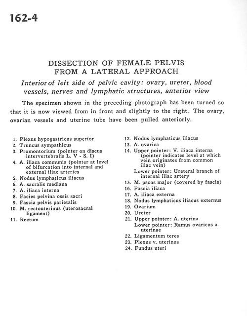
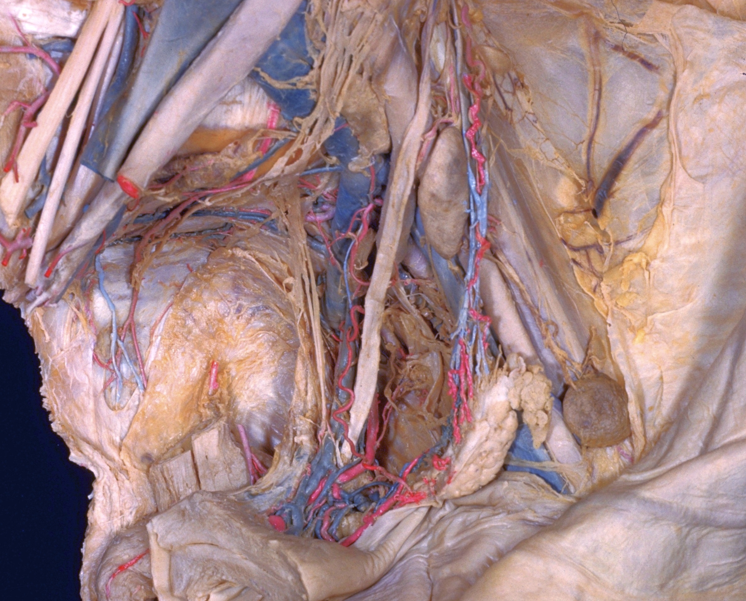
Dissection of female pelvis from a lateral approach
Interior of left side of pelvic cavity.
The specimen shown in the preceding photograph has been turned so that it is now viewed from in front and slightly to the right. The ovary, ovarian vessels and uterine tube have been pulled anteriorly.
- Superior hypogastric plexus
- Sympathetic trunk
- Promontory (pointer on intervertebral disc L. V - S. I)
- Common iliac artery (pointer at level of bifurcation into internal and external iliac arteries)
- Iliac lymph node
- Middle sacral artery
- Internal iliac artery
- Pelvic surface of sacrum
- Parietal pelvic fascia
- Rectouterinus muscle (uterosacral ligament)
- Rectum
- Iliac lymph node
- Ovarian artery
- Upper pointer: Internal iliac vein (pointer indicates level at which vein originates from common iliac vein) Lower pointer: Ureteral branch of internal iliac artery
- Psoas major muscle (covered by fascia)
- Iliac fascia
- External iliac artery
- External iliac lymph node
- Ovary
- Ureter
- Upper pointer: Uterine artery Lower pointer: Ovarian branch of uterine artery
- Ligamentum teres
- Uterine venous plexus
- Fundus of uterus