Dissection of female pelvis from a lateral approach
Uterosacral ligament and cardinal ligament of uterus, medial view
Stanford holds the copyright to the David L. Bassett anatomical images and has assigned
Creative Commons license Attribution-Share
Alike 4.0 International to all of the images.
For additional information regarding use and permissions,
please contact the Medical History Center.
Image #162-2

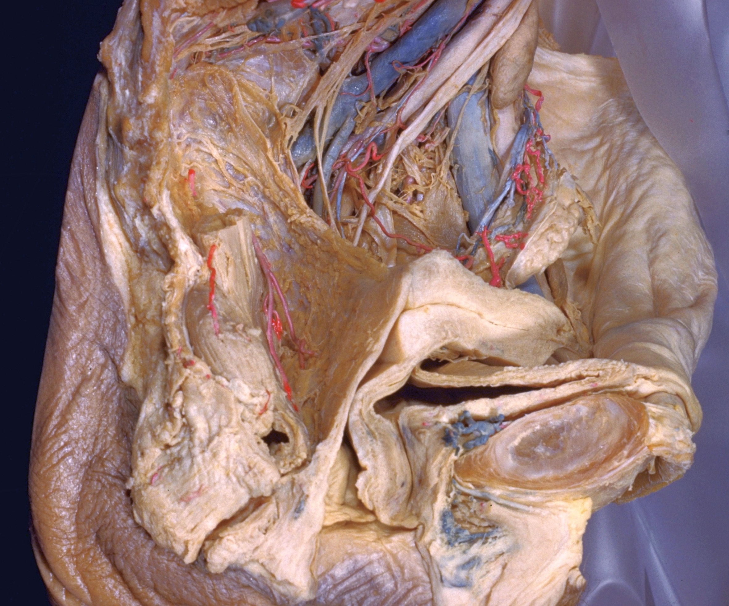
Dissection of female pelvis from a lateral approach
Uterosacral ligament and cardinal ligament of uterus, medial view
The peritoneal covering of the rectouterine fold previously illustrated (161-4) has now been removed. The uterus and vagina have been pulled anteriorly to expose the smooth muscle and fibrous tissue (4) that occupies this fold and extends from the uterus and vaginal wall to the lateral and posterior walls of the pelvic cavity. Although it appears as a continuous sheet, this tissue has usually been subdivided into uterosacral and cardinal (or lateral cervical) ligaments. The former is also identified as the m. rectouterinus and the latter as Mackenrodt's ligament. In subsequent dissections of this series it may be seen that the pelvic plexus of nerves as well as several blood vessels are incorporated within these ligaments.
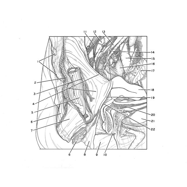
- Coccyx
- Pelvic diaphragm
- Superior rectal artery
- Upper pointer: Rectouterinus muscle (uterosacral ligament) Lower pointer: Cardinal ligament (of Mackenrodt)
- Rectum
- External anal sphincter muscle
- Longitudinal muscle of anal canal
- Central tendon of perineum
- Vestibule of vagina
- External urethral opening
- Superior hypogastric plexus (note that these fibers become incorporated below in the uterosacral ligament)
- Internal iliac vein
- Internal iliac artery
- Ureter
- External iliac vein
- Internal iliac lymph node
- Ovary (retracted anteriorly)
- Uterus
- Posterior wall of vagina
- Urinary bladder
- Pubic symphysis
- Vagina