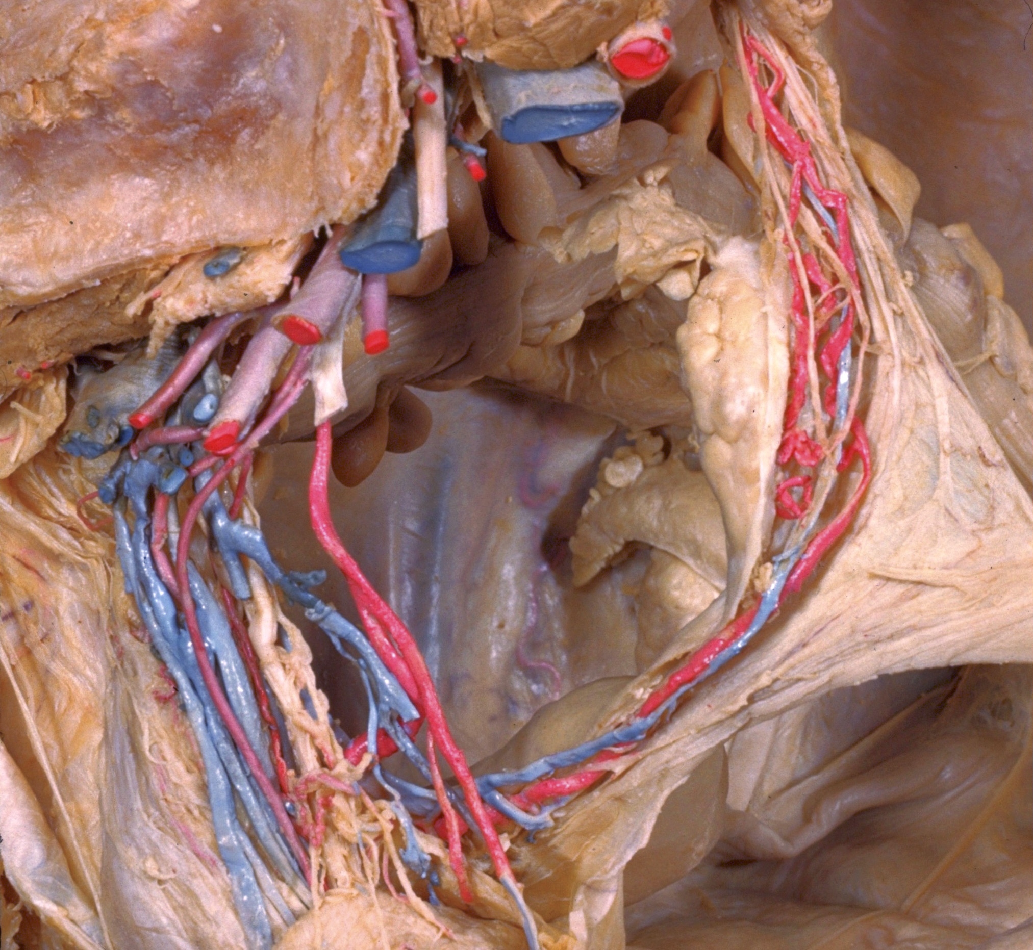
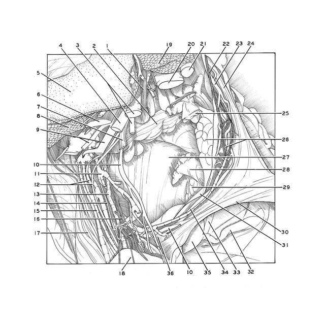
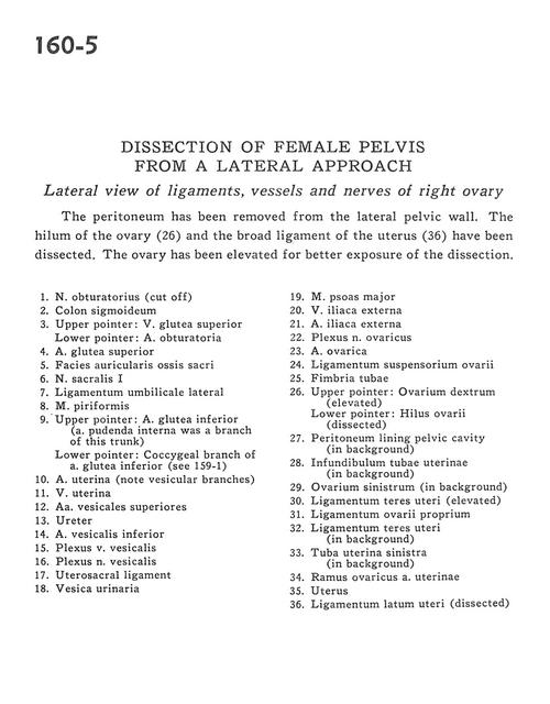
Dissection of female pelvis from a lateral approach
Lateral view of ligaments, vessels and nerves of right ovary
Stanford holds the copyright to the David L. Bassett anatomical images and has assigned
Creative Commons license Attribution-Share
Alike 4.0 International to all of the images.
For additional information regarding use and permissions,
please contact the Medical History Center.
Image #160-5
Dissection of female pelvis from a lateral approach
Lateral view of ligaments, vessels and nerves of right ovary
The peritoneum has been removed from the lateral pelvic wall. The hilum of the ovary (26) and the broad ligament of the uterus (36) have been dissected. The ovary has been elevated for better exposure of the dissection.
- Obturator nerve (cut off)
- Sigmoid colon
- Upper pointer: Superior gluteal vein Lower pointer: Obturator artery
- Superior gluteal artery
- Articular surface of sacrum
- Sacral nerve I
- Lateral umbilical ligament
- Piriform muscle
- Upper pointer: Inferior gluteal artery (internal pudendal artery was a branch of this trunk) Lower pointer: Coccygeal branch of inferior gluteal artery (see 159-1)
- Uterine artery (note vesicular branches)
- Uterine vein
- Superior vesical arteries
- Ureter
- Inferior vesical artery
- Vesical venous plexus
- Vesical nerve plexus
- Uterosacral ligament
- Urinary bladder
- Psoas major muscle
- External iliac vein
- External iliac artery
- Ovarian nerve plexus
- Ovarian artery
- Suspensory ligament of ovary
- Fimbria of uterine tube
- Upper pointer: Right ovary (elevated) Lower pointer: Hilus of ovary (dissected)
- Peritoneum lining pelvic cavity (in background)
- Infundibulum of uterine tube (in background)
- Left ovary (in background)
- Ligamentum teres (of uterus) (elevated)
- Proper ovarian ligament
- Ligamentum teres (of uterus) (in background)
- Left uterine tube (in background)
- Ovarian branch of uterine artery
- Uterus
- Broad ligament of uterus (dissected)