Exploration of the brain from the medial aspect
Internal capsule, ansa lenticularis and subthalamic nucleus
Stanford holds the copyright to the David L. Bassett anatomical images and has assigned
Creative Commons license Attribution-Share
Alike 4.0 International to all of the images.
For additional information regarding use and permissions,
please contact the Medical History Center.
Image #16-7
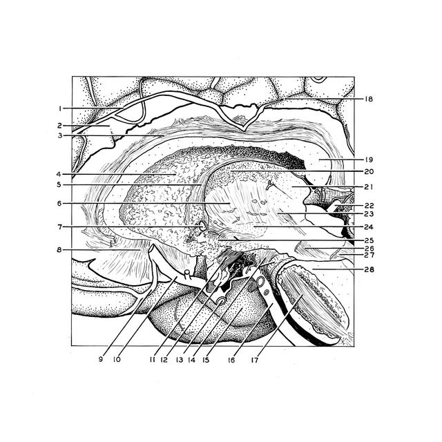
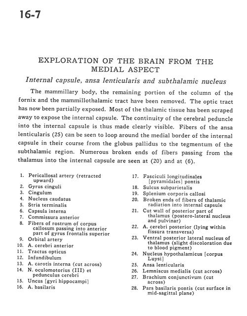
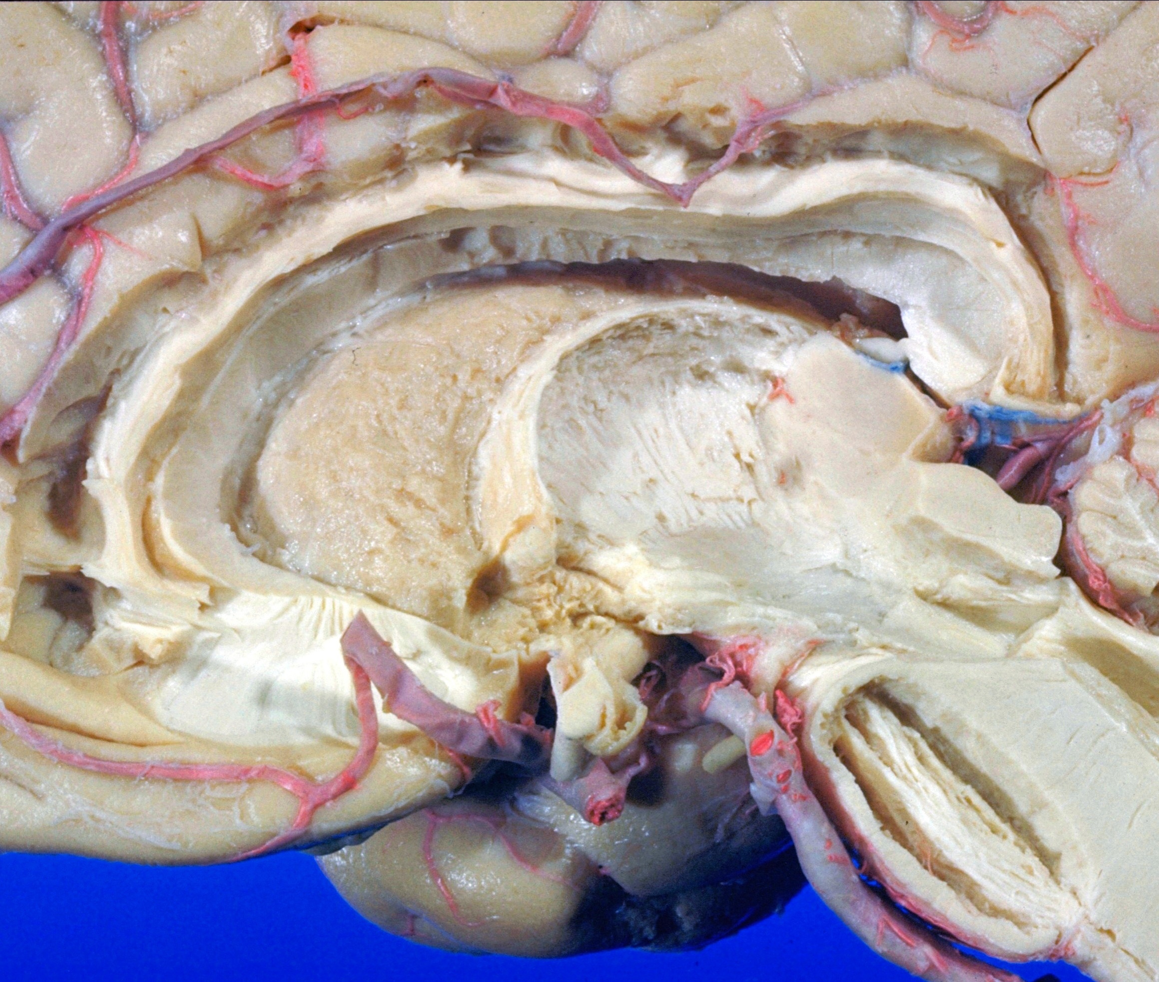
Exploration of the brain from the medial aspect
Internal capsule, ansa lenticularis and subthalamic nucleus
The mammillary body, the remaining portion of the column of the fornix and mammillothalamic tract have been removed. The optic tract has now been partially exposed. Most of the thalamic tissue has been scraped away to expose the internal capsule. The continuity of the cerebral peduncle into the internal capsule is thus made clearly visible. Fibers of the ansa lenticularis (25) can be seen to loop around the medial border of the internal capsule in their course from the globus pallidus to the tegmentum of the subthalamic region. Numerous broken ends of fibers passing from the thalamus into the internal capsule are seen at (20) and at (6).
- Pericallosal artery (retracted upward)
- Cingulate gyrus
- Cingulum
- Caudate nucleus
- Stria terminalis
- Internal capsule
- Anterior commissure
- Fibers of rostrum of corpus callosum passing into anterior part of superior frontal gyrus
- Orbital artery
- Anterior cerebral artery
- Optic tract
- Infundibulum
- Internal carotid artery (cut across)
- Oculomotor nerve (III) and cerebral peduncle
- Uncus (hippocampal gyrus)
- Basilar artery
- Longitudinal fasciculus (pyramidal) of pons
- Subparietal sulcus
- Corpus callosum (splenium)
- Broken ends of fibers of thalamic radiation into internal capsule
- Cut wall of posterior part of thalamus (postero-lateral nucleus and pulvinar)
- Posterior cerebral artery (lying within transverse fissure)
- Ventral posterior lateral nucleus of thalamus (slight discoloration due to blood pigment)
- Hypothalamic nucleus
- Ansa lenticularis
- Medial lemniscus (cut across)
- Brachium conjunctivum (superior cerebellar peduncle) (cut across)
- Basilar part of pons (cut surface in midsagittal plane)