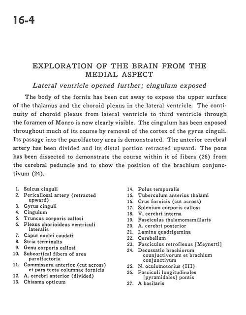
Exploration of the brain from the medial aspect
Lateral ventricle opened further; cingulum exposed
Stanford holds the copyright to the David L. Bassett anatomical images and has assigned
Creative Commons license Attribution-Share
Alike 4.0 International to all of the images.
For additional information regarding use and permissions,
please contact the Medical History Center.
Image #16-4
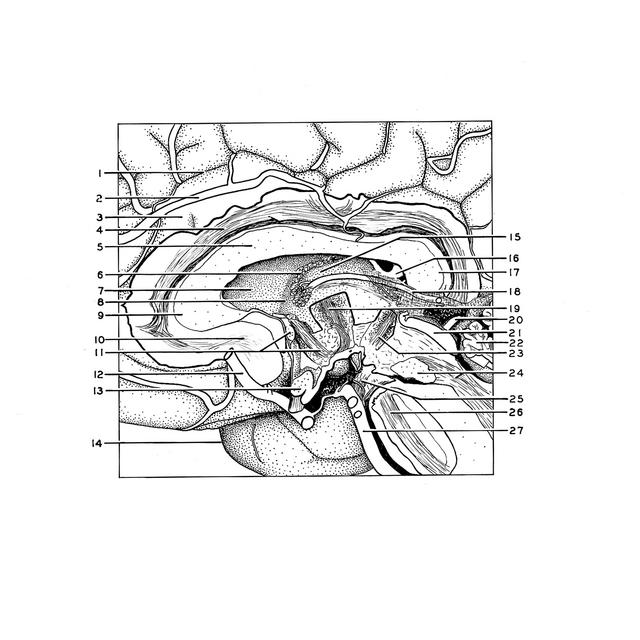
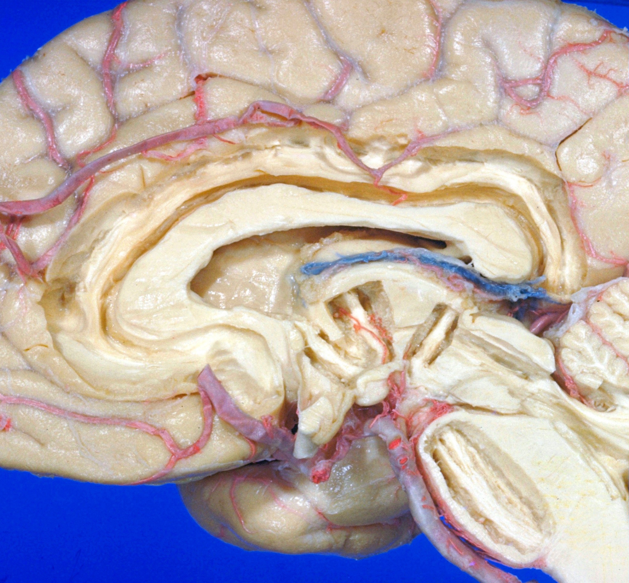
Exploration of the brain from the medial aspect
Lateral ventricle opened further; cingulum exposed
The body of the fornix has been cut away to expose the upper surface of the thalamus and the choroid plexus in the lateral ventricle. The continuity of choroid plexus from lateral ventricle to third ventricle through the foramen of Monro is now clearly visible. The cingulum has been exposed throughout much of its course by removal of the cortex of the gyrus cinguli. Its passage into the paraolfactory area is demonstrated. The anterior cerebral artery has been divided and its distal portion retracted upward. The pons has been to dissected to demonstrate the course within it of fibers (26) from the cerebral peduncle and to show the position of the brachium conjunctivum (24).
- Cingulate sulcus
- Pericallosal artery (retracted upward)
- Cingulate gyrus
- Cingulum
- Corpus callosum (trunk)
- Choroid plexus lateral ventricle
- Head of caudate nucleus
- Stria terminalis
- Genu corpus callosum
- Subcortical fibers of parolfactory area
- Anterior commissure (cut across) and tectal part of column of fornix
- Anterior cerebral artery (divided)
- Optic chiasm
- Temporal pole
- Anterior tubercle of thalamus
- Fornix (crus) (cut across)
- Corpus callosum (splenium)
- Internal cerebral vein
- Mamillothalamic tract
- Posterior cerebral artery
- Quadrigeminal plate
- Cerebellum
- Fasciculus retroflexus
- Decussation brachium conjuctivum and brachium conjunctivum (superior cerebellar peduncle)
- Oculomotor nerve (III)
- Longitudinal fasciculus (pyramidal of pons)
- Basilar artery