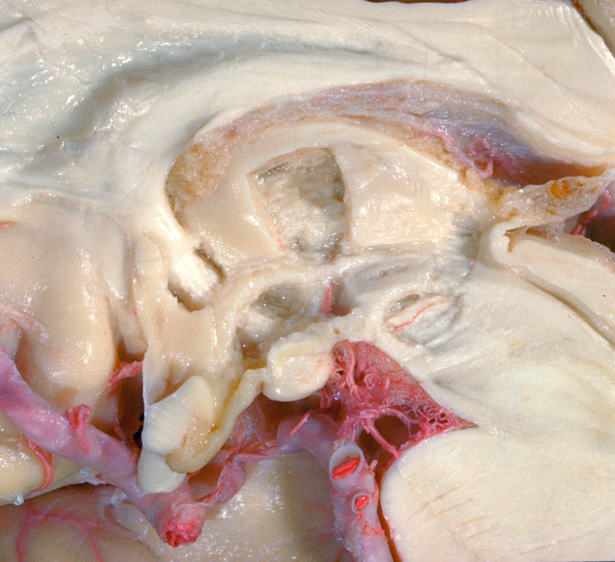
Exploration of the brain from the medial aspect
Column of fornix, mammillothalamic tract and periventricular fibers
Stanford holds the copyright to the David L. Bassett anatomical images and has assigned
Creative Commons license Attribution-Share
Alike 4.0 International to all of the images.
For additional information regarding use and permissions,
please contact the Medical History Center.
Image #16-1
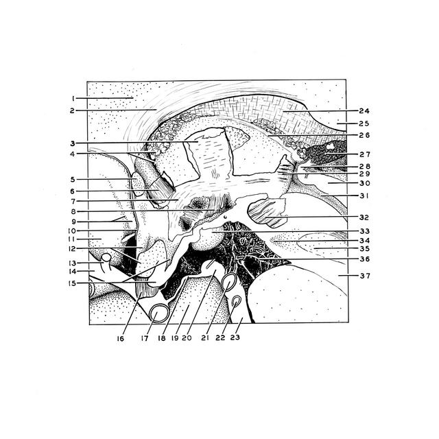
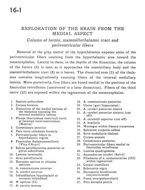
Exploration of the brain from the medial aspect
Column of fornix, mammillothalamic tract and periventricular fibers
Removal of the gray matter of the hypothalamus exposes some of the periventricular fibers coursing from the hypothalamic area toward the mesencephalon. Lateral to these, in the depths of the dissection, the column of the fornix (6) is seen as it approaches the mammillary body and the mammillothalamic tract (8) as it leaves. The dissected area (3) of the thalamus contains longitudinally coursing fibers of the internal medullary lamina. More posteriorly, fine fibers are found medial to the position of the fasciculus retroflexus (uncovered in a later dissection). Fibers of the third nerve (32) are exposed within the tegmentum of the mesencephalon.
- Septum pellucidum
- Fornix (body)
- Dissection of the medial nucleus of the thalamus exposing the internal medullary lamina
- Choroid plexus third ventricle and interventricular foramen
- Anterior commissure
- Tectal part of column of fornix
- Periventricular fibers in hypothalamic region
- Mamillothalamic tract
- Posterior parolfactory sulcus and subcallosal gyrus
- Lamina terminalis
- Parolfactory area
- Optic recess and optic chiasm
- Anterior communicating artery
- Anterior cerebral artery
- Infundibulum of hypothalamus and infundibular recess
- Optic nerve (II)
- Internal carotid artery
- Posterior communicating artery
- Uncus (hippocampal gyrus)
- Posterior cerebral artery right
- Posterior cerebral artery left (cut off)
- Superior cerebellar artery (cut oft)
- Basilar artery
- Meninges within transverse fissure
- Corpus callosum (splenium)
- Stria medullaris thalami
- Pineal body
- Posterior commissure
- Periventricular fibers medial to fasciculus retroflexus
- Quadrigeminal plate
- Cerebral aqueduct
- Filaments of oculomotor nerve (III) within tegmentum
- Mamillary body
- Substantia nigra
- Decussation brachium conjunctivum
- Interpeduncular fossa
- Dorsal part of pons