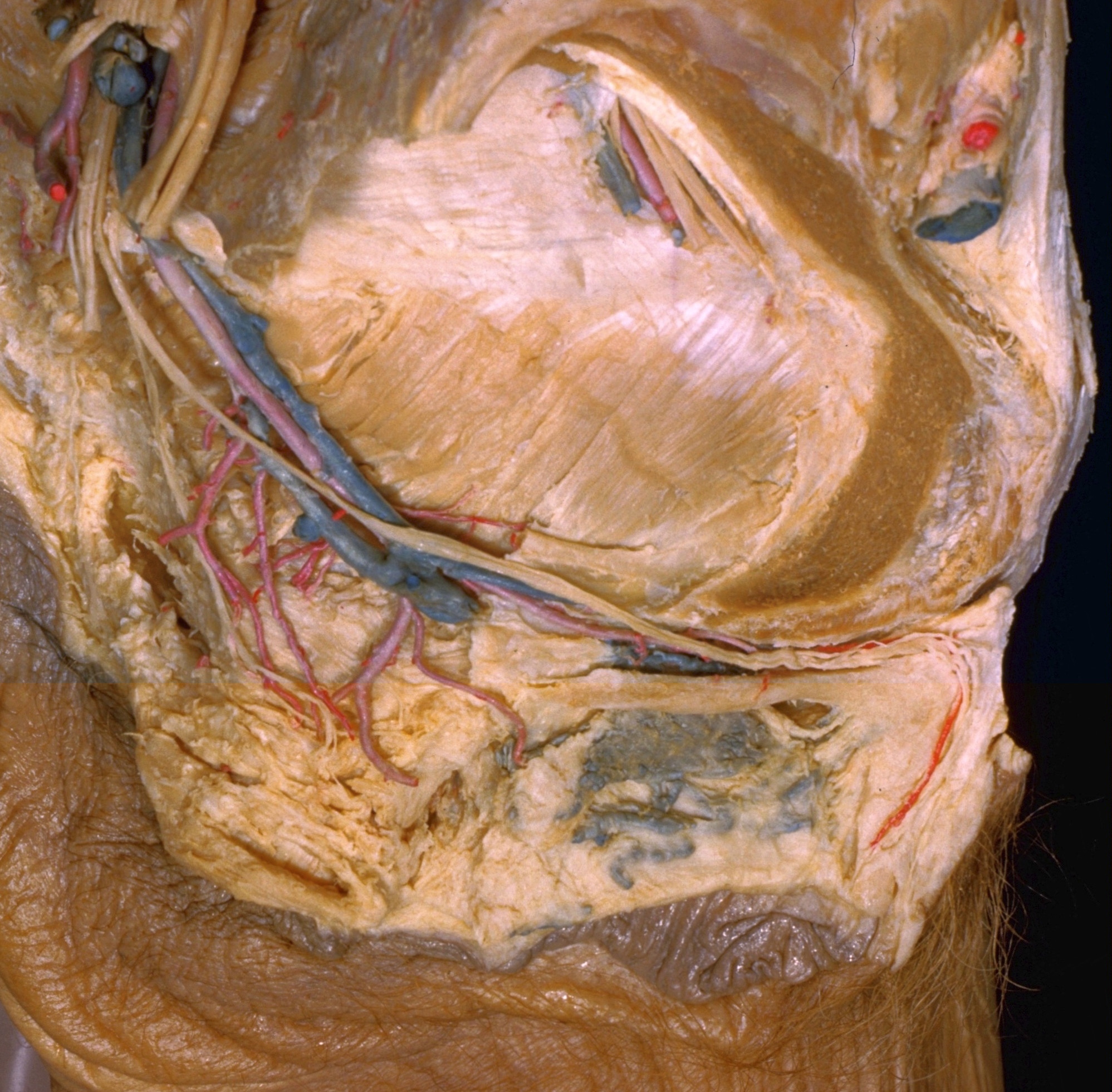
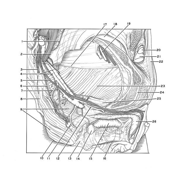
Dissection of female pelvis froma lateral approach
Pelvic diaphragm, lateral view with pudendal vessels and nerves in situ
Stanford holds the copyright to the David L. Bassett anatomical images and has assigned
Creative Commons license Attribution-Share
Alike 4.0 International to all of the images.
For additional information regarding use and permissions,
please contact the Medical History Center.
Image #159-4
Dissection of female pelvis froma lateral approach
Pelvic diaphragm, lateral view with pudendal vessels and nerves in situ
In an earlier view of this specimen (158-7) the pelvic diaphragm was partially visible through a window cut in the lateral pelvic wall. The exposure of the diaphragm has been increased by resection of more of the ischium. The pudendal vessels and nerves have been retained in place, although the fascia (25) that formed the walls of the pudendal canal has been nearly completely removed from around them.
- Greater sciatic notch
- Ischial spine
- Inferior gluteal nerve (cut off)
- Internal pudendal artery and vein
- Pudendal nerve
- Inferior rectal nerve
- Inferior rectal artery
- Anococcygeal ligament
- External anal sphincter muscle
- Anus
- Perineal artery
- Dorsal artery of clitoris
- Central tendon of perineum
- Greater vestibular (Bartholin's) gland
- Vestibular bulb
- Vestibule of vagina
- Tendinous arch of levator ani muscle (atypical, see discussion of 158-7)
- Acetabulum (remnant)
- Obturator artery
- Femoral artery
- Femoral vein
- Inguinal ligament
- Levator ani muscle
- Body of pubic bone (partially cut away)
- Obturator fascia (forming medial wall of pudendal canal)
- Body of clitoris