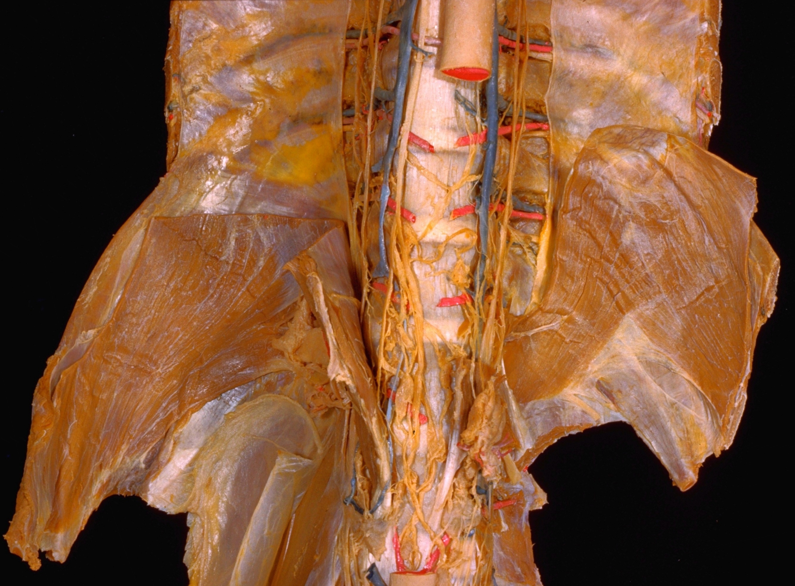
Kidneys, suprarenal glands and posterior abdominal vessels, nerves and glands
Thoracic duct, abdominal and lower thoracic parts viewed from in front
Stanford holds the copyright to the David L. Bassett anatomical images and has assigned
Creative Commons license Attribution-Share
Alike 4.0 International to all of the images.
For additional information regarding use and permissions,
please contact the Medical History Center.
Image #152-4
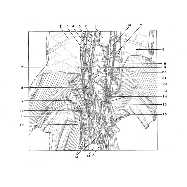
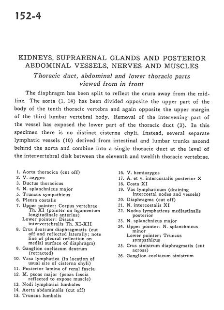
Kidneys, suprarenal glands and posterior abdominal vessels, nerves and glands
Thoracic duct, abdominal and lower thoracic parts viewed from in front
The diaphragm has been split to reflect the crura away from the midline. The aorta (1,14) has been divided opposite the upper part of the body of the tenth thoracic vertebra and again opposite the upper margin of the third lumbar vertebral body. Removal of the intervening part of the vessel has exposed the lower part of the thoracic duct (3). In this specimen there is no distinct cisterna chylli. Instead, several separate lymphatic vessels (10) derived from intestinal and lumbar trunks ascend behind the aorta and combine into a single thoracic duct at the level of intervertebral disk between the eleventh and twelfth thoracic vertebrae.
- Thoracic aorta (cut off)
- Azygos vein
- Thoracic duct
- Greater splanchnic nerve
- Sympathetic trunk
- Costal pleura
- Upper pointer: Body of vertebra Th. XI (pointer on anterior longitudinal ligament) Lower pointer: Intervertebral disc Th. XI-XII
- Right crus of diaphragm (cut off and reflected laterally note line of pleural reflection on medial surface of diaphragm)
- Right celiac ganglion (retracted)
- Lymph vessel (in location of usual site of cisterna chyli)
- Posterior lamina of renal fascia
- Psoas major muscle (psoas fascia reflected to expose muscle)
- Lumbar lymph nodes
- Abdominal aorta (cut off)
- Lumbar trunk
- Hemiazygos vein
- Posterior intercostal artery and vein X
- Rib XI
- Lymph vessel (draining intercostal nodes and vessels)
- Diaphragm (cut off)
- Intercostal nerve XI
- Posterior mediastinal lymph node
- Greater splanchnic nerve
- Upper pointer: Lesser splanchnic nerve Lower pointer: Sympathetic trunk
- Left crus of diaphragm (cut across)
- Left celiac ganglion