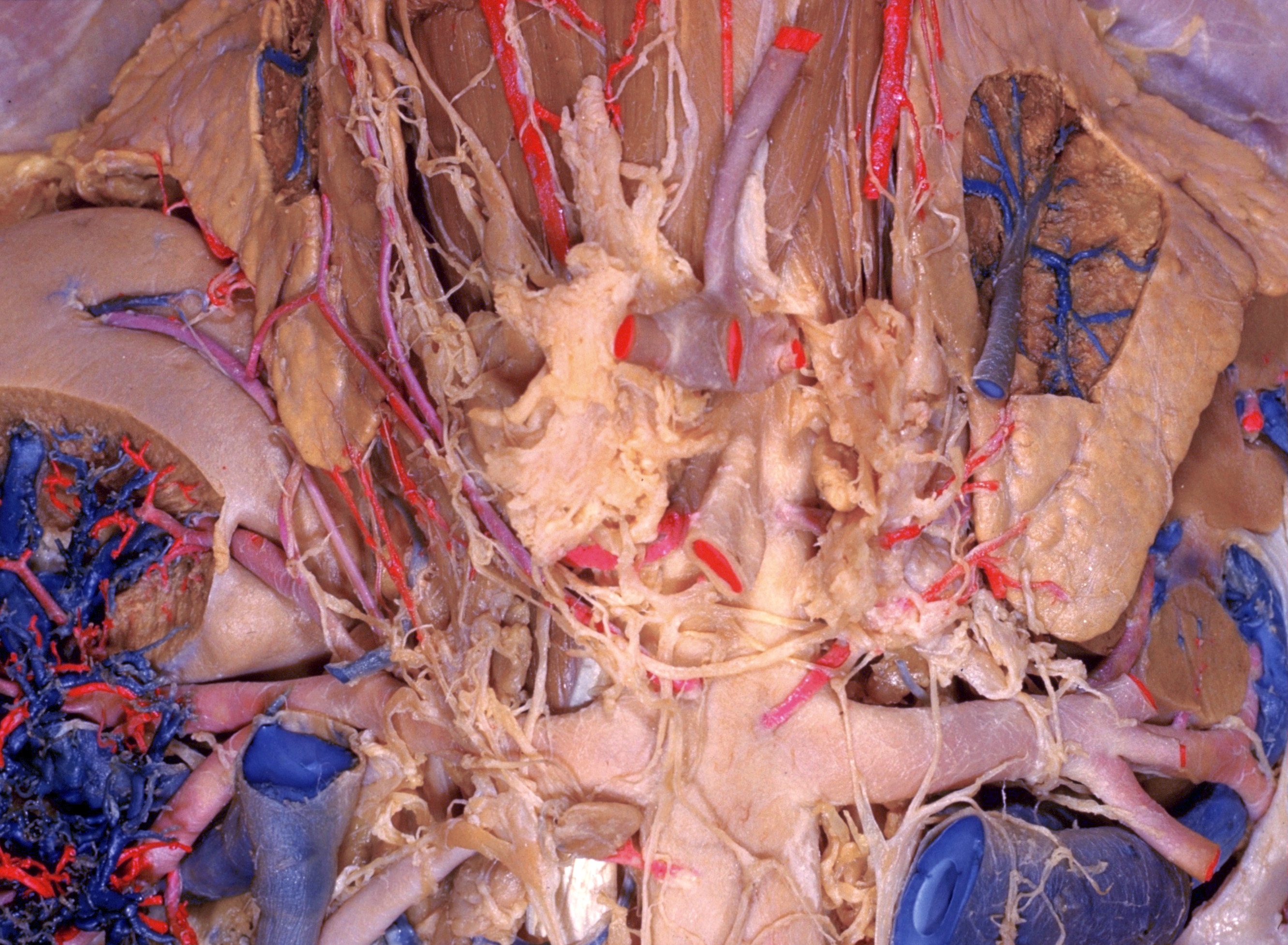
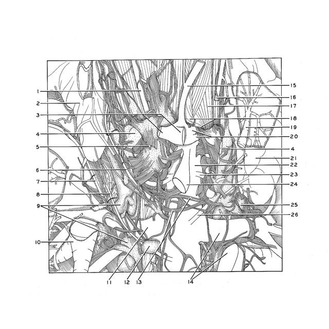
Kidneys, suprarenal glands and posterior abdominal vessels, nerves and muscles
Close-up view of celiac plexus, celiac ganglia, and origins of celiac and superior mesenteric arteries
Stanford holds the copyright to the David L. Bassett anatomical images and has assigned
Creative Commons license Attribution-Share
Alike 4.0 International to all of the images.
For additional information regarding use and permissions,
please contact the Medical History Center.
Image #151-2
Kidneys, suprarenal glands and posterior abdominal vessels, nerves and muscles
Close-up view of celiac plexus, celiac ganglia, and origins of celiac and superior mesenteric arteries
The plexus surrounding the celiac and superior mesenteric arteries has been split and retracted to the side to reveal the origins of these arteries from the aorta. The emergence of the aorta through the aortic haitus of the diaphragm is partly visible. The continuity of the celiac, superior mesenteric, aorticorenal and aortic plexuses is apparent in this specimen.
- Right inferior phrenic artery
- Right suprarenal gland
- Left pointer: Common hepatic artery (cut off) Right pointer: Splenic artery (cut off)
- Celiac ganglion
- Celiac plexus (right and left sides divided longitudinally)
- Middle suprarenal artery
- Polar artery to right kidney (source of some inferior suprarenal arteries see previous views)
- Inferior suprarenal artery (branch of right renal artery)
- Aorticorenal ganglion
- Right renal vein (cut off near attachment to inferior vena cava)
- Right renal artery
- Lumbar lymph node (lateral aortic node)
- Interconnecting fibers between autonomic plexuses of two sides
- Left renal artery and vein
- Left gastric artery
- Left inferior phrenic artery (cut off)
- Diaphragm
- Upper pointer: Suspensory muscle of duodenum (cut off) Lower pointer: Margin of aortic hiatus
- Left suprarenal vein
- Upper pointer: Celiac trunk Lower pointer: Left inferior phrenic artery (cut off)
- Left suprarenal gland
- Abdominal aorta
- Middle suprarenal artery
- Superior mesenteric artery
- Left aorticorenal ganglion
- Inferior suprarenal artery