Kidneys, suprarenal glands and posterior abdominal vessels, nerves and muscles
Blood supply and nerves of left suprarenal gland, close-up view
Stanford holds the copyright to the David L. Bassett anatomical images and has assigned
Creative Commons license Attribution-Share
Alike 4.0 International to all of the images.
For additional information regarding use and permissions,
please contact the Medical History Center.
Image #150-7
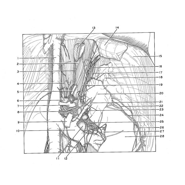
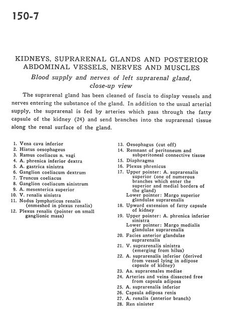
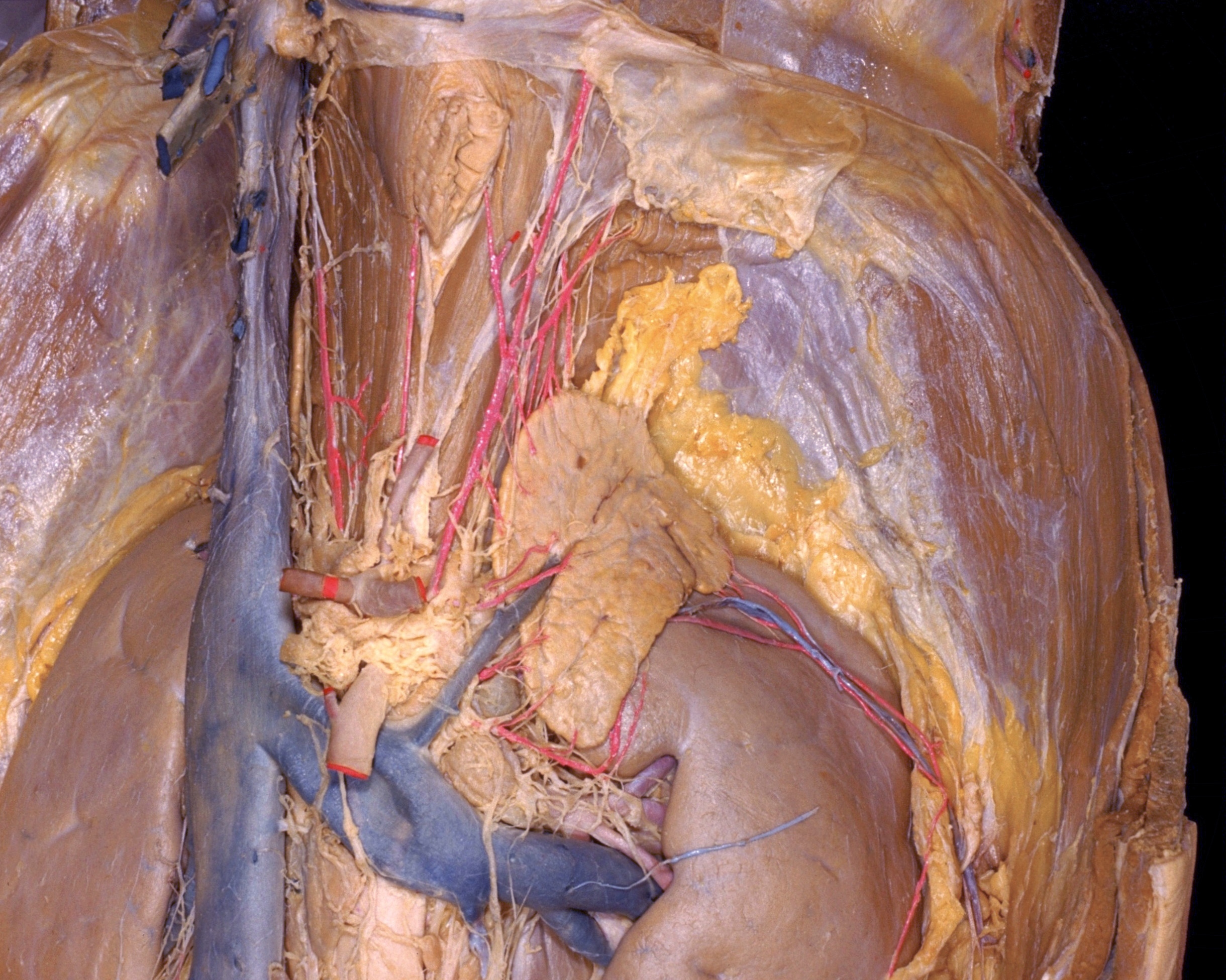
Kidneys, suprarenal glands and posterior abdominal vessels, nerves and muscles
Blood supply and nerves of left suprarenal gland, close-up view
The suprarenal gland has been cleaned of fascia to display vessels and nerves entering the substance of the gland. In addition to the usual arterial supply, the suprarenal is fed by arteries which pass through the fatty capsule of the kidney (24) and send branches into the suprarenal tissue along the renal surface of the gland.
- Inferior vena cava
- Esophageal hiatus
- Celiac branch of vagus nerve
- Right inferior phrenic artery
- Left gastric artery
- Right celiac ganglion
- Celiac trunk
- Left celiac ganglion
- Superior mesenteric artery
- Left renal vein
- Renal lymph node (enmeshed in renal plexus)
- Renal plexus (pointer on small ganglionic mass)
- Esophagus (cut off)
- Remnant of peritoneum and subperitoneal connective tissue
- Diaphragm
- Phrenic plexus
- Upper pointer: Superior suprarenal artery (one of numerous branches which enter the superior and medial borders of the gland) Lower pointer: Superior margin of suprarenal gland
- Upward extension of fatty capsule of kidney
- Upper pointer: Left inferior phrenic artery Lower pointer: Medial margin of suprarenal gland
- Anterior surface of suprarenal gland
- Left suprarenal vein (emerging from hilus)
- Inferior suprarenal artery (derived from vessel lying in fatty capsule of kidney)
- Middle suprarenal arteries
- Arteries and veins dissected free from fatty capsule
- Inferior suprarenal artery
- Fatty capsule of kidney
- Renal artery (anterior branch)
- Left kidney