Kidneys, suprarenal glands and posterior abdominal vessels, nerves and muscles
Blood supply and nerves of right suprarenal gland, close-up view
Stanford holds the copyright to the David L. Bassett anatomical images and has assigned
Creative Commons license Attribution-Share
Alike 4.0 International to all of the images.
For additional information regarding use and permissions,
please contact the Medical History Center.
Image #150-3
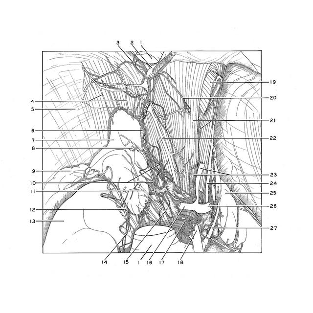
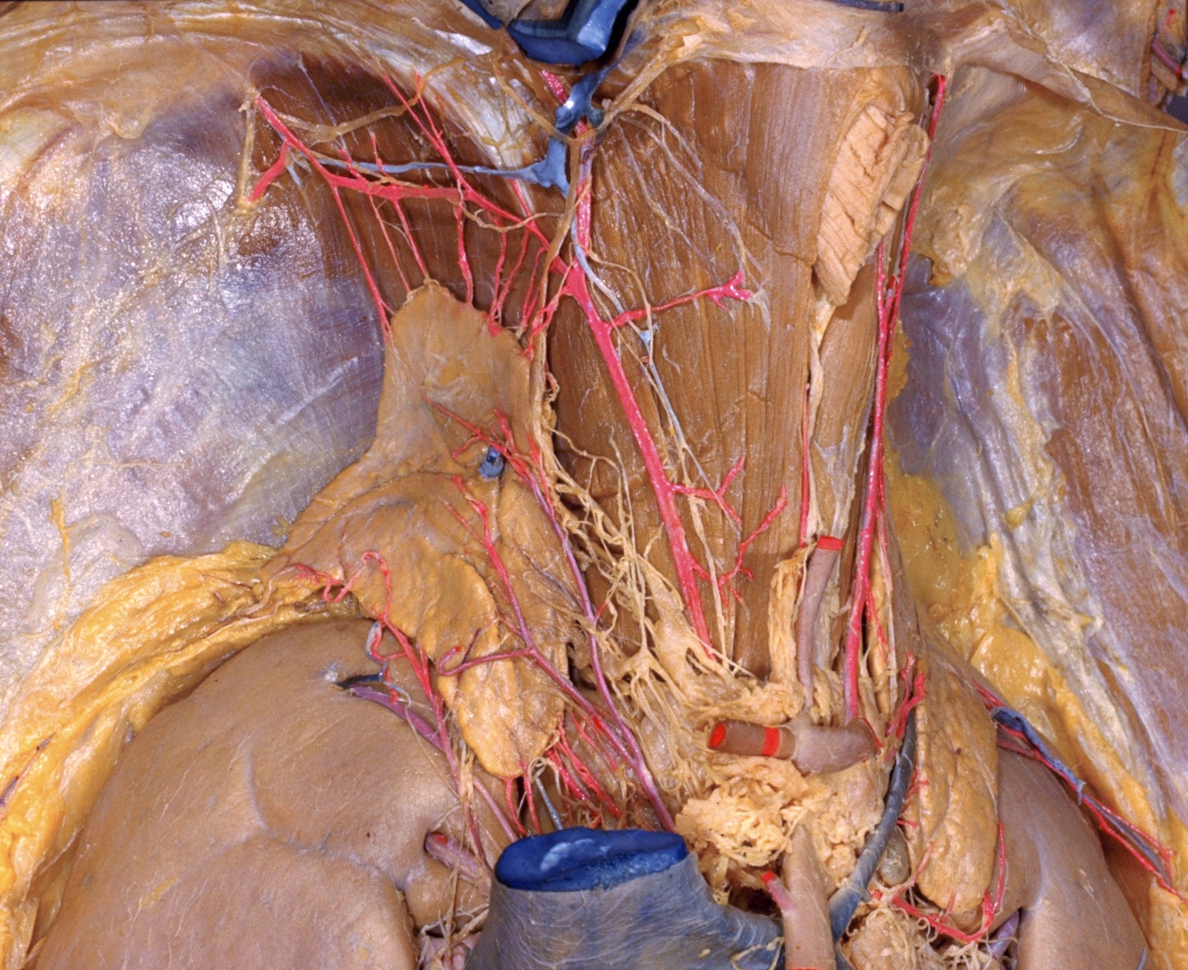
Kidneys, suprarenal glands and posterior abdominal vessels, nerves and muscles
Blood supply and nerves of right suprarenal gland, close-up view
The inferior vena cava (1) has been removed from the central field of the dissection. Vessels, nerves and ganglia related to the suprarenal glands have been exposed. A large communication (6) occurs between the coeliac plexus and the right phrenic nerve. The plexiform arrangement of nerves on the inferior surface of the diaphragm has also been displayed.
- Inferior vena cava (divided)
- Inferior phrenic vein
- Right phrenic nerve
- Superior suprarenal arteries
- Diaphragm
- Communicating nerve between celiac plexus and inferior phrenic plexus (pointer on phrenic ganglion)
- Right suprarenal gland
- Right suprarenal vein (cut off at point of entry into vena cava)
- Pararenal fat
- Suprarenal plexus
- Middle suprarenal artery (origin from aorta visible in next view)
- Inferior suprarenal artery
- Right kidney
- Branch of renal artery
- Celiac ganglion
- Common hepatic artery (cut off)
- Celiac plexus
- Superior mesenteric artery
- Esophagus
- Branch of right phrenic nerve to crus of diaphragm
- Upper pointer: Right crus of diaphragm Lower pointer: Celiac branch of vagus nerve
- Right inferior phrenic artery
- Upper pointer: Left gastric artery Lower pointer: Left gastric plexus
- Left inferior phrenic artery
- Left suprarenal gland
- Splenic artery
- Left suprarenal vein