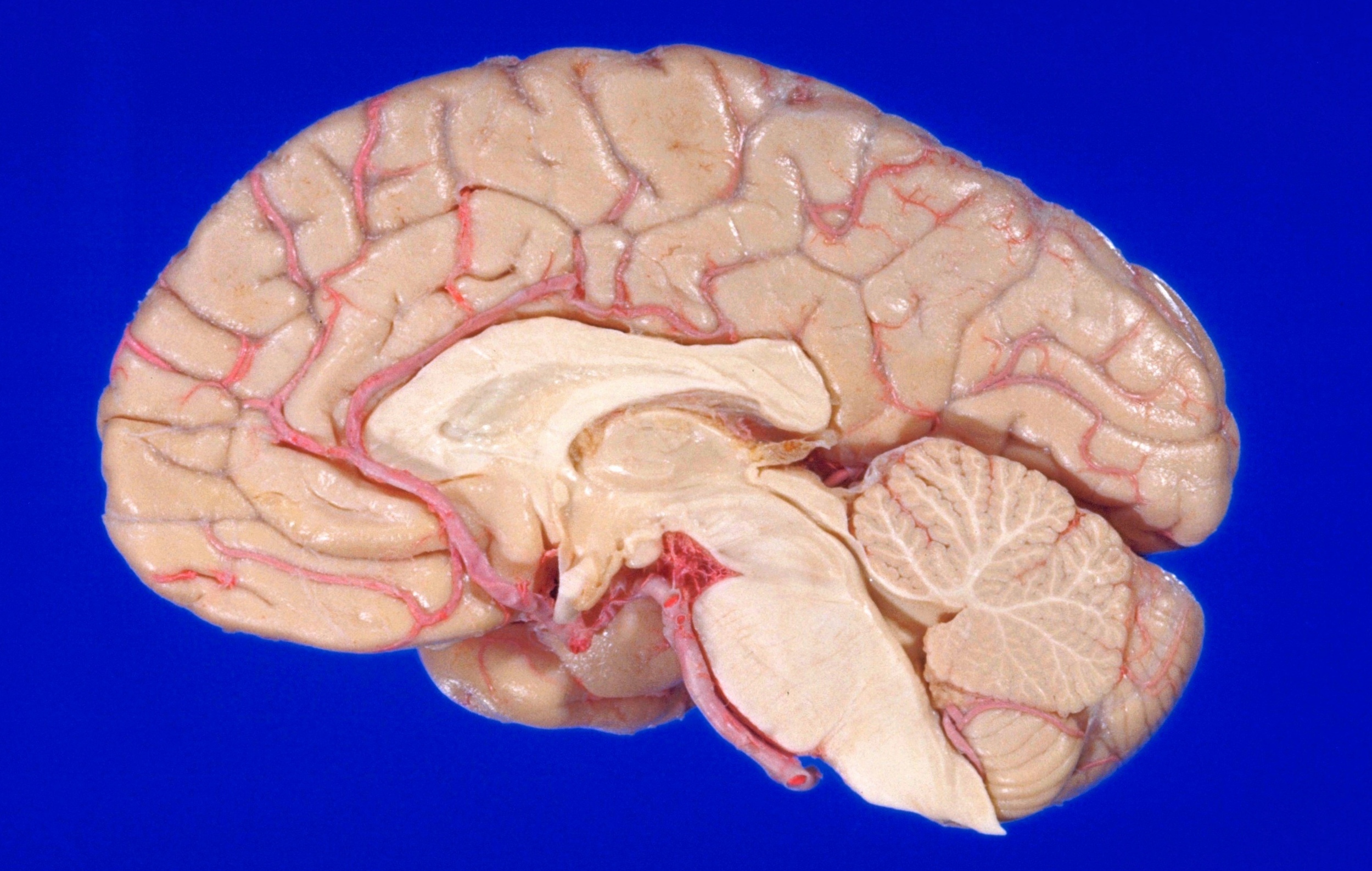
Exploration of the brain from the medial aspect
Median sagittal section
Stanford holds the copyright to the David L. Bassett anatomical images and has assigned
Creative Commons license Attribution-Share
Alike 4.0 International to all of the images.
For additional information regarding use and permissions,
please contact the Medical History Center.
Image #15-6

Exploration of the brain from the medial aspect
Median sagittal section
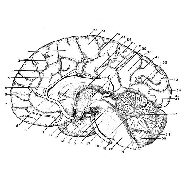
The brain is divided in the mid-sagittal plane to expose the medial surface of the cerebral hemisphere and midline structures of the brain stem and cerebellum. The third ventricle, cerebral aqueduct, fourth ventricle and central canal are exposed. In the telencephalon the actual, cut surfaces include the corpus callosum, rostral lamina, lamina terminalis, septum pellucidum, fornix and anterior commissure. In the diencephalon the structures divided are the optic chiasma, infundibulum and floor of the third ventricle, massa intermedia, tela chorioidea of the roof of the third ventricle, pineal body and the habenular and posterior commissures. The hypophysis has been removed. The basilar artery is intact, but its branches to the left are all divided. The distribution of the anterior cerebral artery to the hemisphere is shown.
- Superior frontal gyrus
- Anterior medial frontal arteries
- Cingulate sulcus (callosomarginal artery lies within this sulcus)
- Cingulate gyrus
- Pericallosal artery
- Frontopolar artery
- Frontal pole
- Genu corpus callosum
- Septum pellucidum and cavit of septum pellucidum
- Orbital artery
- Anterior communicating artery (cut off)
- Optic chiasm
- Internal carotid artery
- Uncus (hippocampal gyrus)
- Cerebral aqueduct
- Pons
- Basilar artery
- Fourth ventricle
- Vertebral artery left (cut off)
- Medulla oblongata
- Central canal
- Central sulcus (of Rolando)
- Paracentral lobule
- Middle medial frontal arteries
- Marginal part cingulate sulcus
- Posterior medial frontal artery
- Fornix (body)
- Subparietal sulcus
- Thalamus, massa intermedia and pineal body
- Precuneus
- Parieto-occipital fissure
- Branch of posterior cerebral artery in calcarine fissure
- Cuneus
- Occipital pole
- Lingual gyrus
- Transverse fissure
- Cerebellum
- Posterior inferior cerebellar artery
- Cerebellar tonsil