Exploration of the brain from its lateral aspect
Lateral ventricle, stria terminalis and internal capsule
Stanford holds the copyright to the David L. Bassett anatomical images and has assigned
Creative Commons license Attribution-Share
Alike 4.0 International to all of the images.
For additional information regarding use and permissions,
please contact the Medical History Center.
Image #15-2
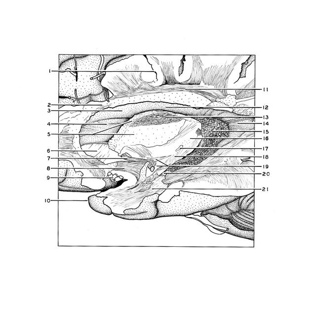
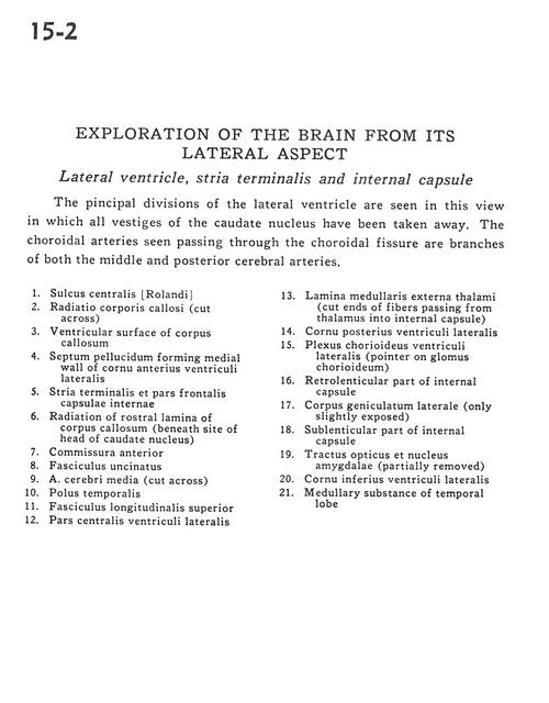
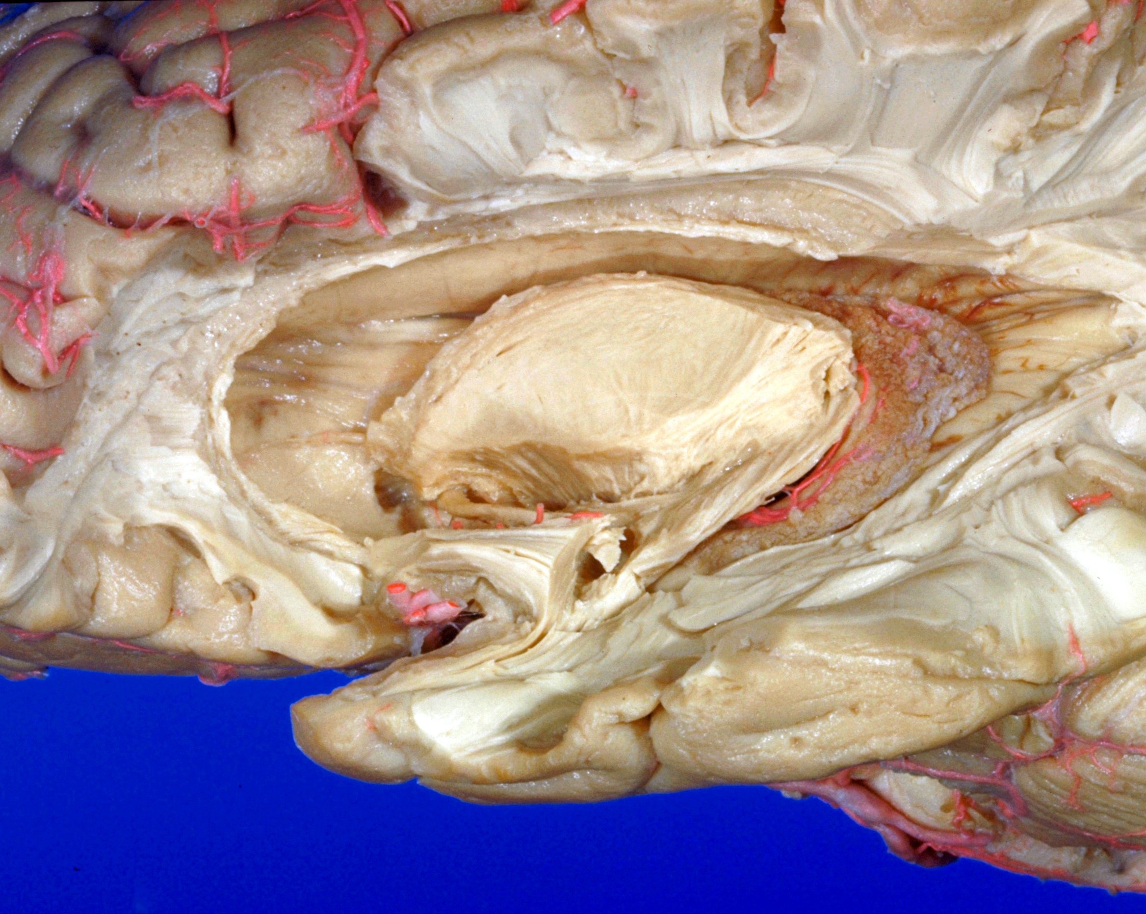
Exploration of the brain from its lateral aspect
Lateral ventricle, stria terminalis and internal capsule
The principal divisions of the lateral ventricle are seen in this view in which all vestiges of the caudate nucleus have been taken away. The choroidal arteries seen passing through the choroidal fissure are branches of both the middle and posterior cerebral arteries.
- Central sulcus (of Rolando)
- Radiations of corpus callosum (cut across)
- Ventricular surface of corpus callosum
- Septum pellucidum forming medial wall of anterior horn lateral ventricle
- Stria terminalis and frontal part internal capsule
- Radiation of rostral lamina of corpus callosum (beneath site of head of caudate nucleus)
- Anterior commissure
- Uncinate fasciculus
- Middle cerebral artery (cut across)
- Temporal pole
- Superior longitudinal fasciculus
- Central part lateral ventricle
- External medullary lamina (thalamus) (cut ends of fibers passing from thalamus into internal capsule)
- Posterior horn lateral ventricle
- Choroid plexus lateral ventricle (pointer on glomus chorioideum)
- Retrolenticular part of internal capsule
- Lateral geniculate body (only slightly exposed)
- Sublenticular part of internal capsule
- Optic tract and amygdaloid nucleus (partially removed)
- Inferior horn of lateral ventricle
- Medullary substance of temporal lobe