Kidneys, suprarenal, glands and posterior abdominal vessels, nerves and muscles
Fascial relations of right kidney, close-up anterior view
Stanford holds the copyright to the David L. Bassett anatomical images and has assigned
Creative Commons license Attribution-Share
Alike 4.0 International to all of the images.
For additional information regarding use and permissions,
please contact the Medical History Center.
Image #148-6
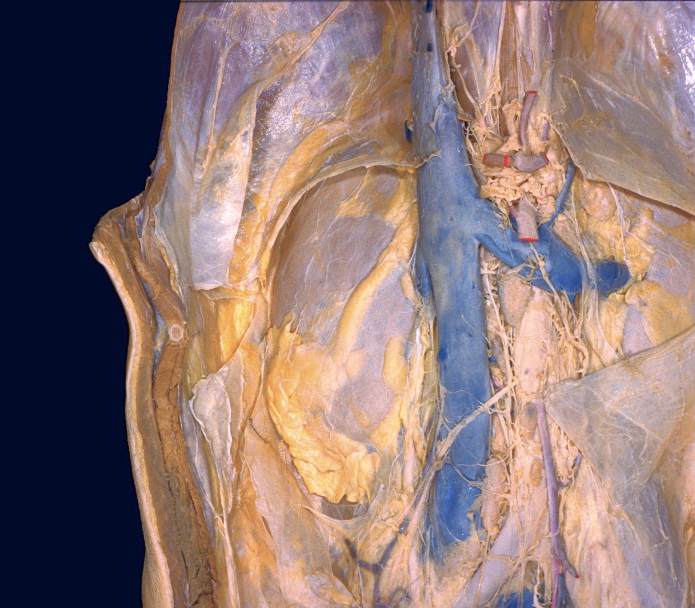
Kidneys, suprarenal, glands and posterior abdominal vessels, nerves and muscles
Fascial relations of right kidney, close-up anterior view
It was possible to separate the renal fascia from the subperitoneal connective tissue in this specimen for a distance of several centimeters lateral to the border of the kidney. Near the lateral border of the kidney this layer splits (6) to pass in front of and behind the kidney. In addition, a fascial lamina (7, middle pointer) passes parallel to the surface of the kidney to interconnect the anterior and posterior layers of renal fascia. This lamina obscures the angular cleft between diverging fascial layers which would otherwise be apparent if the renal fascia were opened and its interior inspected following removal of the kidney.
- Right suprarenal gland (covered by fascia)
- Hepatic vein from lower part of right lobe of liver (cut off)
- Peritoneum (medial part elevated)
- Kidney (covered by connective tissue of fatty capsule)
- Anterior layer of renal fascia (elevated)
- Lateral confluence of anterior layer with posterior lamina of renal fascia
- Upper and lower pointers: Renal fascia (extension anterolaterally external to peritoneum) Middle pointer: Connecting layer between anterior and posterior laminae of renal fascia
- Transversalis fascia
- Fatty capsule
- Fascial lamina passing medially from anterior layer of renal fascia below lower pole of kidney
- Crus of diaphragm
- Celiac trunk
- Celiac ganglion
- Superior mesenteric artery
- Left renal vein
- Right renal vein
- Lamina of fascia related to adipose capsule of left kidney
- Inferior vena cava
- Abdominal aorta
- Right testicular vein (lower path of this vein indicated by brownish discoloration instead of blue latex)
- Testicular artery (uncolored)
- Inferior mesenteric artery
- Filament of testicular plexus
- Filament of aortic plexus
- Lumbar lymph node