Kidneys, suprarenal, glands and posterior abdominal vessels, nerves and muscles
Renal fascia, general anterior view
Stanford holds the copyright to the David L. Bassett anatomical images and has assigned
Creative Commons license Attribution-Share
Alike 4.0 International to all of the images.
For additional information regarding use and permissions,
please contact the Medical History Center.
Image #148-5
Kidneys, suprarenal, glands and posterior abdominal vessels, nerves and muscles
Renal fascia, general anterior view
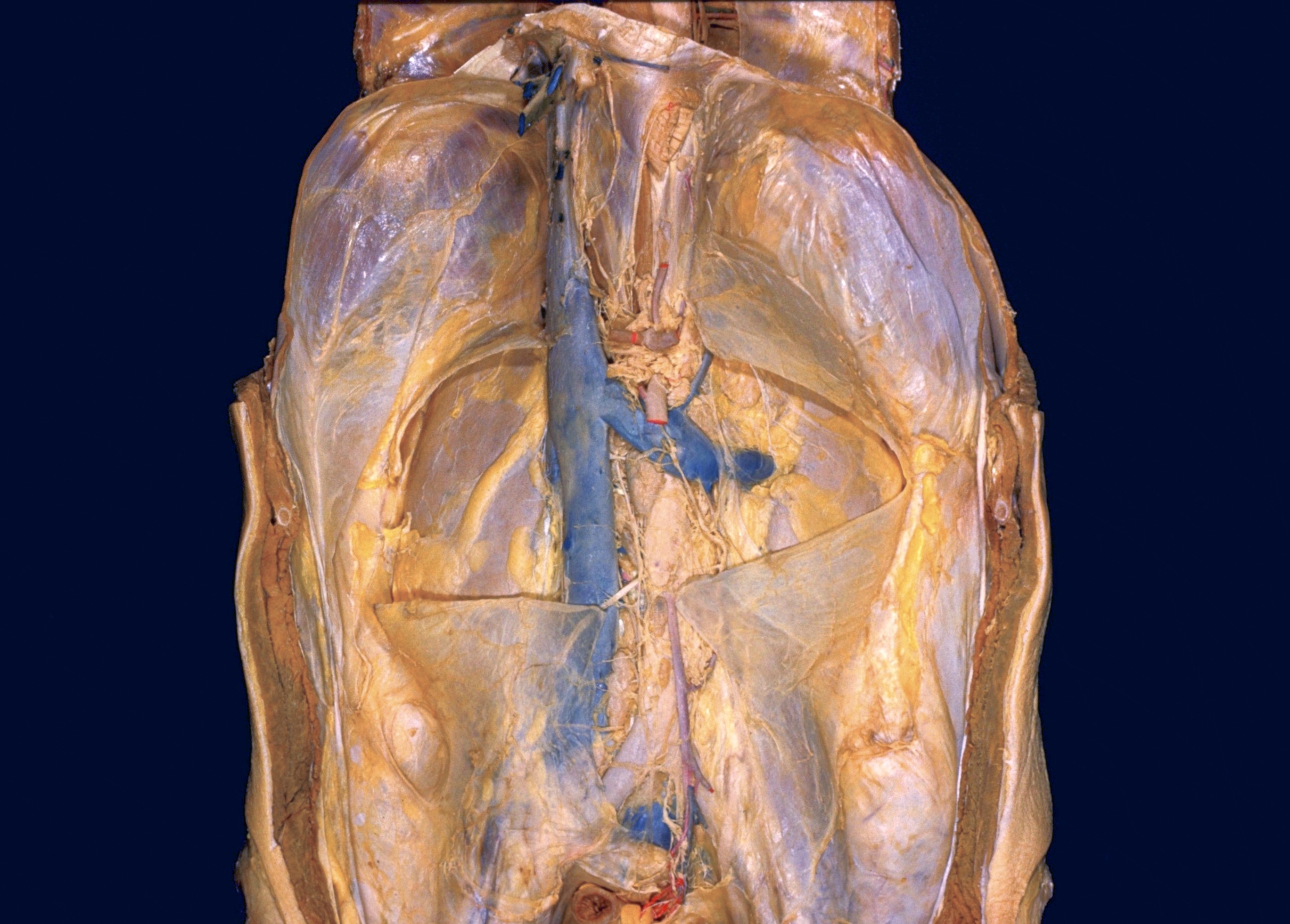
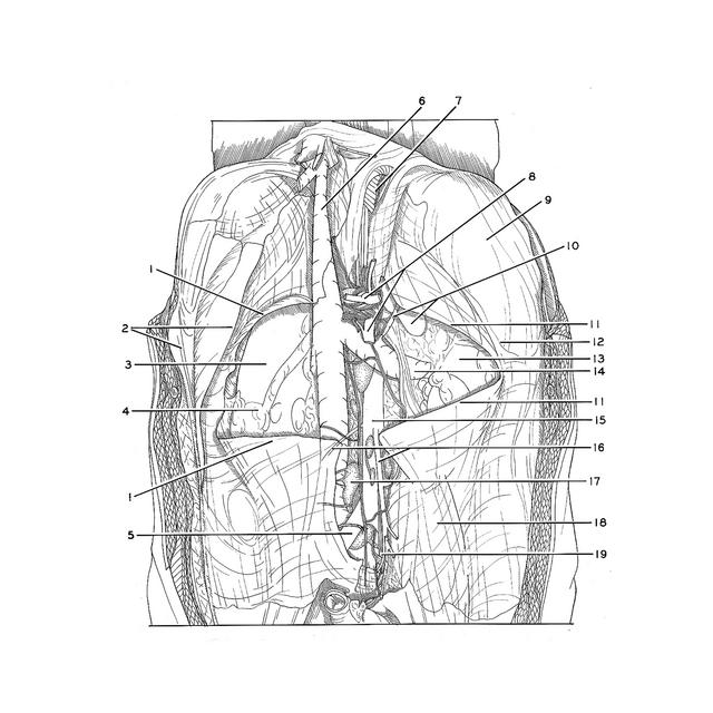
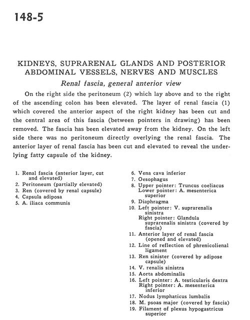
On the right side the peritoneum (2) which lay above and to the right of the ascending colon has been elevated. The layer of renal fascia (1) which covered the anterior aspect of the right kidney has been cut and the central area of this fascia (between pointers in drawing) has been removed. The fascia has been elevated away from the kidney. On the left side there was no peritoneum directly overlying the renal fascia. The anterior layer of renal fascia has been cut and elevated to reveal the underlying fatty capsule of the kidney.
- Renal fascia (anterior layer, cut and elevated)
- Peritoneum (partially elevated)
- Kidney (covered by renal capsule)
- Fat capsule
- Common iliac artery
- Inferior vena cava
- Esophagus
- Upper pointer: Celiac trunk Lower pointer: Superior mesenteric artery
- Diaphragm
- Left pointer: Left suprarenal vein Right pointer: Left suprarenal gland (covered by fascia)
- Anterior layer of renal fascia (opened and elevated)
- Line of reflection of phrenicosplenic ligament
- Left kidney (covered by adipose capsule)
- Left renal vein
- Abdominal aorta
- Left pointer: Right testicular artery Right pointer: Inferior mesenteric artery
- Lumbar lymph node
- Psoas major muscle (covered by fascia)
- Filament of superior hypogastric plexus