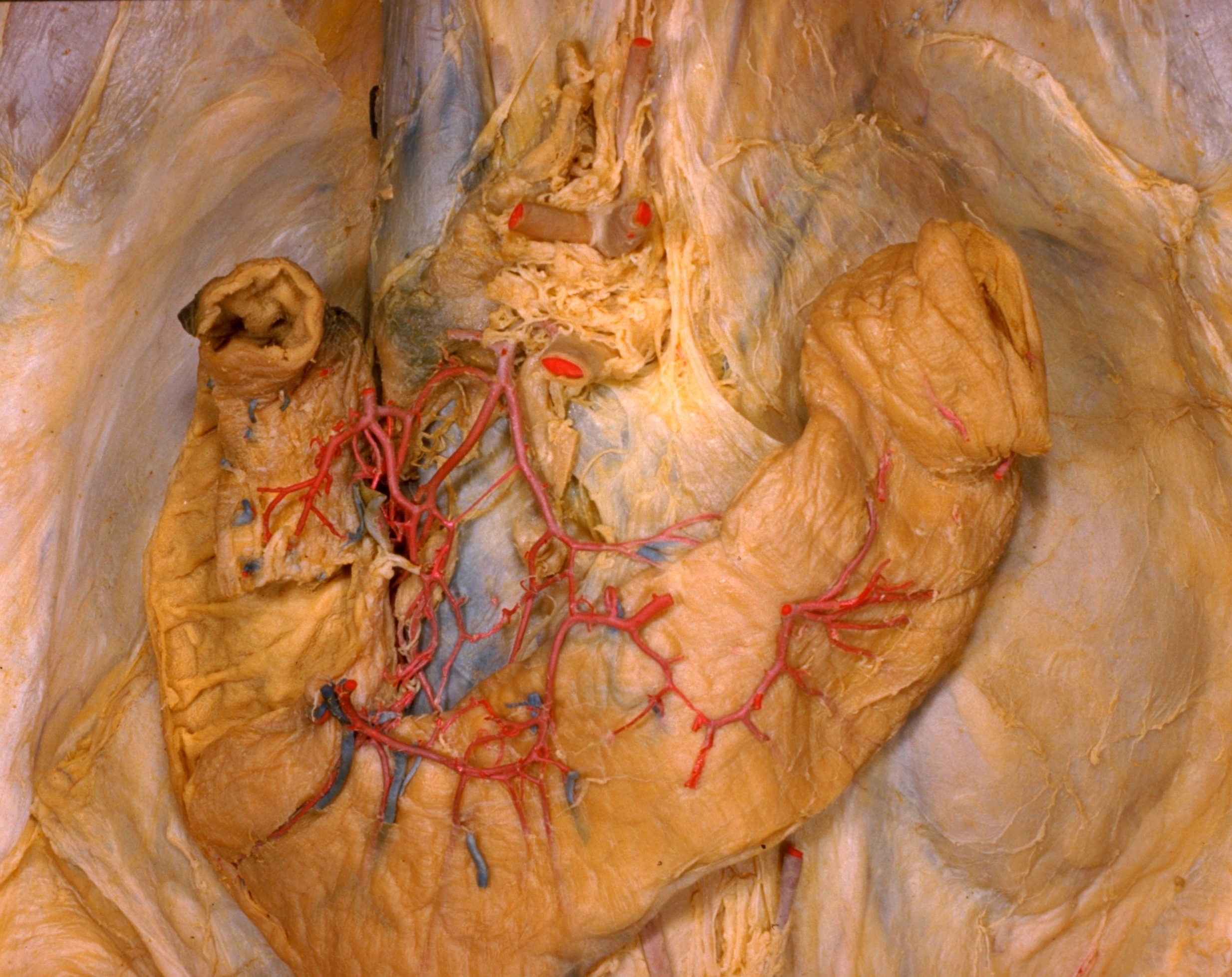
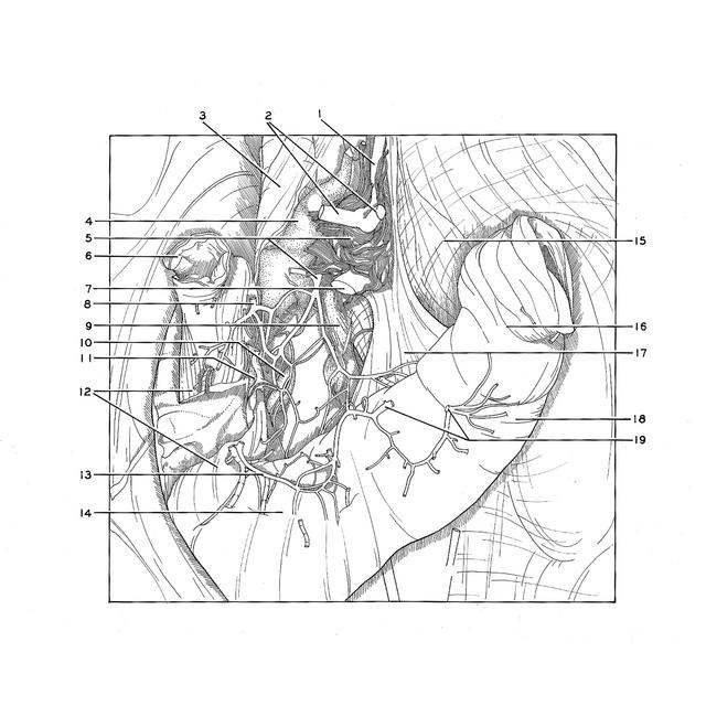
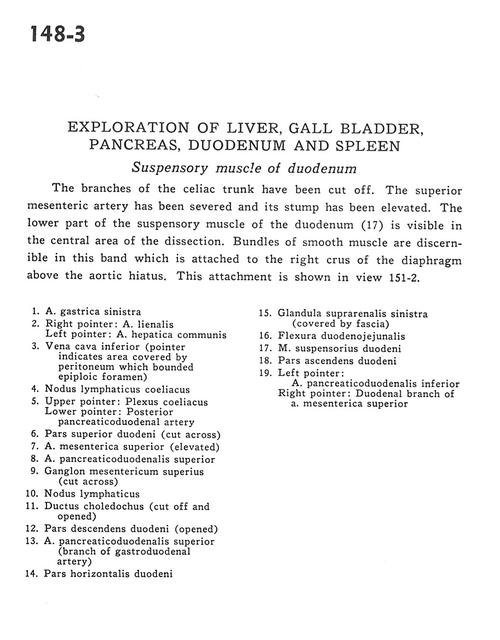
Exploration of liver, gall bladder, pancreas, duodenum and spleen
Suspensory muscle of duodenum
Stanford holds the copyright to the David L. Bassett anatomical images and has assigned
Creative Commons license Attribution-Share
Alike 4.0 International to all of the images.
For additional information regarding use and permissions,
please contact the Medical History Center.
Image #148-3
Exploration of liver, gall bladder, pancreas, duodenum and spleen
Suspensory muscle of duodenum
The branches of the cellac trunk have been cut off. The superior mesenteric artery has been severed and its stump has been elevated.The lower part of the suspensory muscle of the duodenum (17) is visible in the central area of the dissection. Bundles of smooth muscle are discernible in this band which is attached to the right crus of the diaphragm above the aortic hiatus. This attachment is shown in view 151-2.
- Left gastric artery
- Right pointer: Splenic artery Left pointer: Common hepatic artery
- Inferior vena cava (pointer indicates area covered by peritoneum which bounded epiploic foramen)
- Celiac lymph node
- Upper pointer: Celiac plexus Lower poInter: Posterior pancreaticoduodenal artery
- Superior part of duodenum (cut across)
- Superior mesenteric artery (elevated)
- Superior pancreaticoduodenal artery
- Superior mesenteric ganglion (cut across)
- Lymph node
- Common bile duct (cut off and opened)
- Descending part of duodenum (opened)
- Superior pancreaticoduodenal artery (branch of gastroduodenal artery)
- Horizontal part of duodenum
- Left suprarenal gland (covered by fascia)
- Duodenojejunal flexure
- Suspensory ligament of duodenum
- Ascending part of duodenum
- Left pointer: Inferior pancreaticoduodenal artery Right pointer: Duodenal branch of superior mesenteric artery