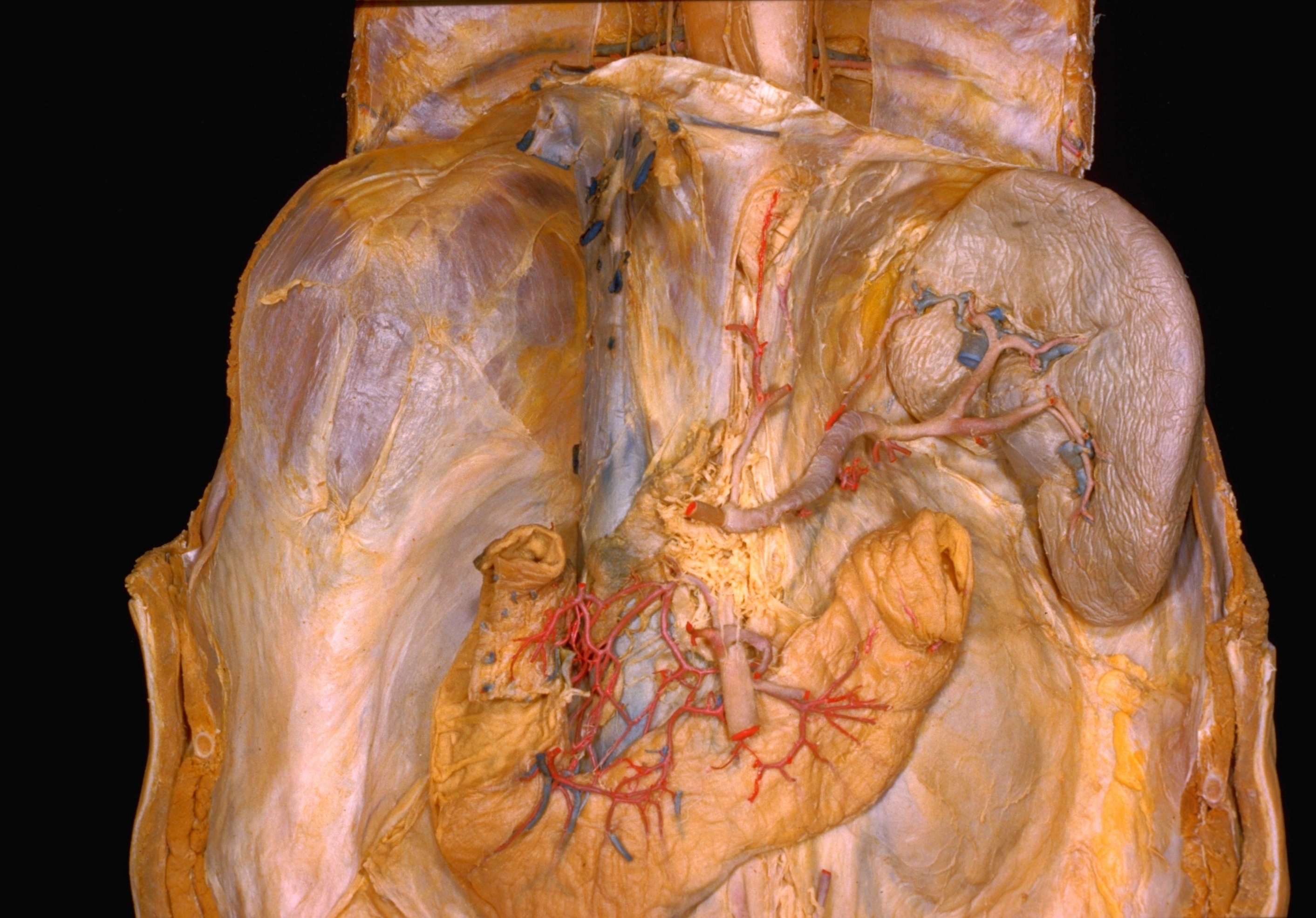
Exploration of liver, gall bladder, pancreas, duodenum and spleen
Spleen and splenic artery
Stanford holds the copyright to the David L. Bassett anatomical images and has assigned
Creative Commons license Attribution-Share
Alike 4.0 International to all of the images.
For additional information regarding use and permissions,
please contact the Medical History Center.
Image #148-2
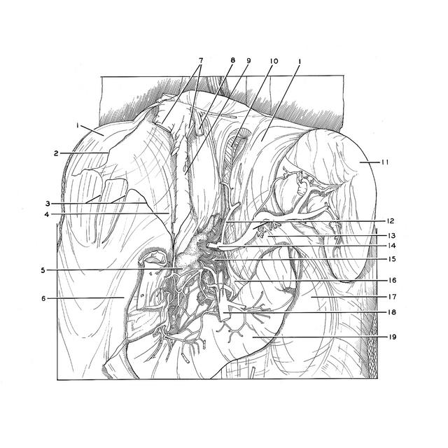
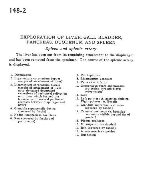
Exploration of liver, gall bladder, pancreas, duodenum and spleen
Spleen and splenic artery
The liver has been cut from its remaining attachment to the diaphragm and has been removed from the specimen. The course of the splenic artery is displayed.
- Diaphragm
- Coronary ligament (upper margin of attachment of liver)
- Coronary ligament (lower margin of attachment of liver; note elongated downward extensions of peritoneal reflection onto liver which formed the boundaries of several peritoneal recesses between diaphragm and liver)
- Right suprarenal gland (covered by fascia)
- Celiac lymph node
- Kidney (covered by fascia and peritoneum)
- Hepatic veins
- Ligamentum venosum
- Inferior vena cava
- Esophagus (abdominal part, projecting through esophageal hiatus)
- Spleen
- Left pointer: Left gastric artery Right pointer: Splenic artery
- Left suprarenal gland (covered by fascia)
- Celiac trunk (common hepatic artery visible beyond tip of pointer)
- Celiac plexus
- Suspensory muscle of duodenum
- Kidney (covered by fascia)
- Superior mesenteric artery
- Duodenum