Exploration of liver, gall bladder, pancreas, duodenum and spleen
Relations of inferior vena cava and liver
Stanford holds the copyright to the David L. Bassett anatomical images and has assigned
Creative Commons license Attribution-Share
Alike 4.0 International to all of the images.
For additional information regarding use and permissions,
please contact the Medical History Center.
Image #147-7
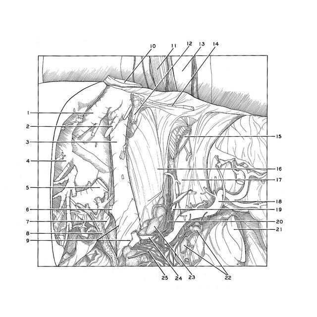
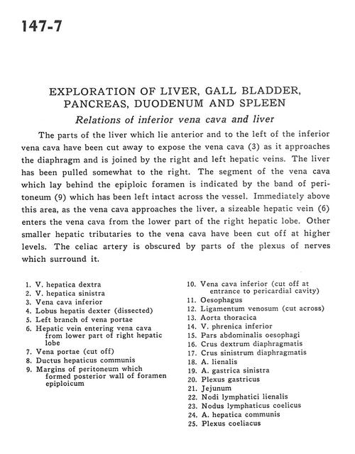
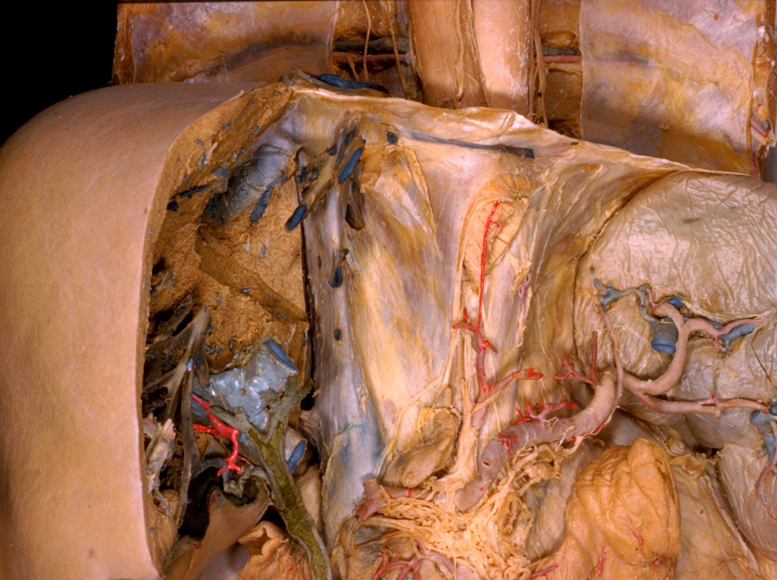
Exploration of liver, gall bladder, pancreas, duodenum and spleen
Relations of inferior vena cava and liver
The parts of the liver which lie anterior and to the left of the inferior vena cava have been cut away to expose the vena cava (3) as it approaches the diaphragm and is joined by the right and left hepatic veins. The liver has been pulled somewhat to the right. The segment of the vena cava which lay behind the epiploic foramen is indicated by the band of peritoneum(9) which has been left intact across the vessel. Immediately above this area, as vena cava approaches the liver, a sizable hepatic vein (6) enters the vena cava from the lower part of the right hepatic lobe. Other smaller hepatic tributaries to the vena cava have been cut off at higher levels. The celiac artery is obscured by parts of the plexus of nerves which surround it.
- Right hepatic vein
- Left hepatic vein
- Inferior vena cava
- Right lobe of liver (dissected)
- Left branch of portal vein
- Hepatic vein entering vena cava from lower part of right hepatic lobe
- Portal vein (cut off)
- Common hepatic duct
- Margins of peritoneum which formed posterior wall of epiploic foramen
- Inferior vena cava (cut off at entrance to pericardial cavity)
- Esophagus
- Ligamentum venosum (cut across)
- Thoracic aorta
- Inferior phrenic vein
- Abdominal part of esophagus
- Right crus of diaphragm
- Left crus of diaphragm
- Splenic artery
- Left gastric artery
- Gastric plexus
- Jejunum
- Splenic lymph nodes
- Celiac lymph node
- Common hepatic artery
- Celiac plexus