Exploration of liver, gall bladder, pancreas, duodenum and spleen
Dissection of duodenum (continued).
Stanford holds the copyright to the David L. Bassett anatomical images and has assigned
Creative Commons license Attribution-Share
Alike 4.0 International to all of the images.
For additional information regarding use and permissions,
please contact the Medical History Center.
Image #147-4
Exploration of liver, gall bladder, pancreas, duodenum and spleen
Dissection of duodenum (continued).
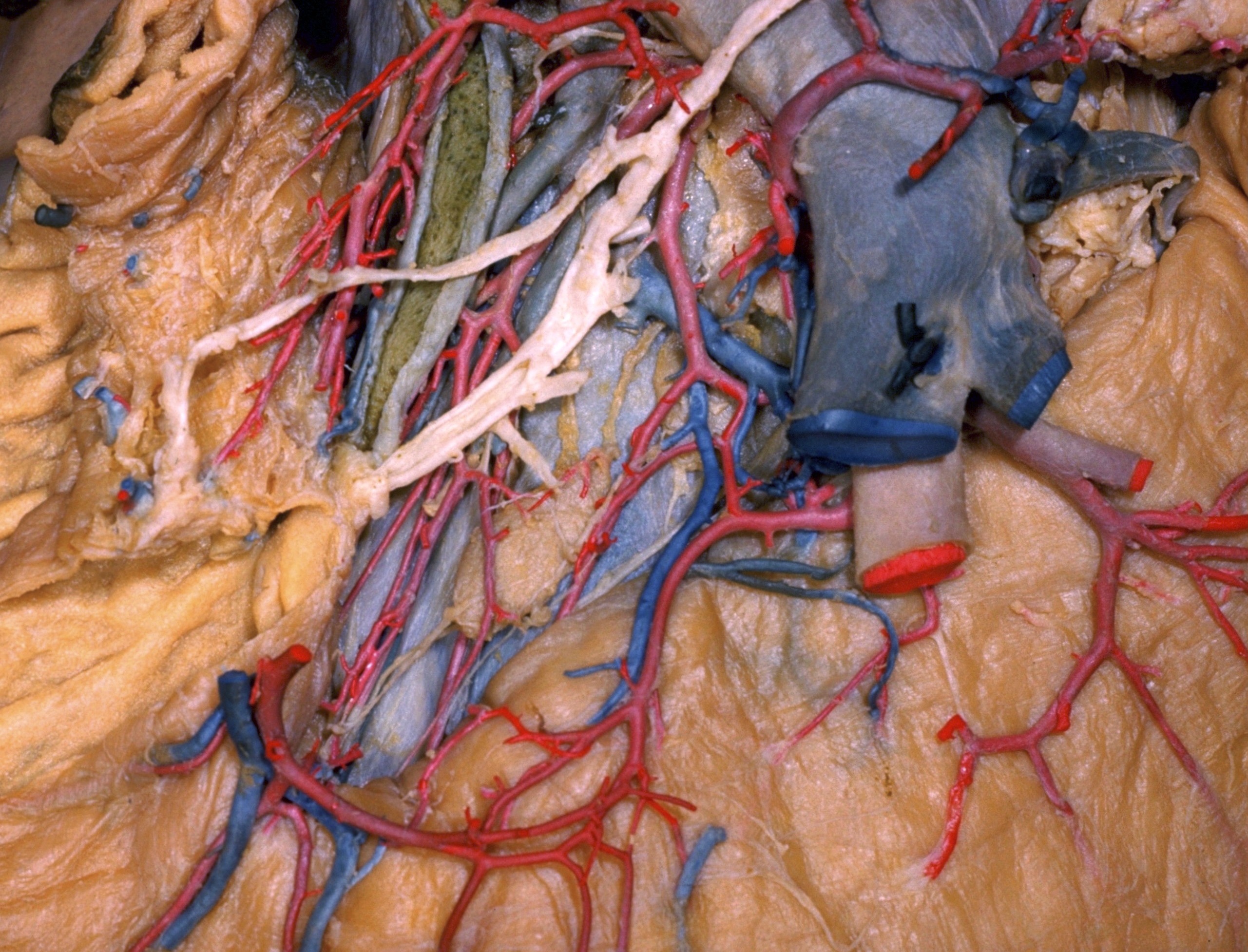
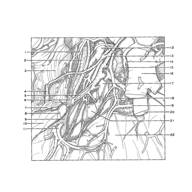
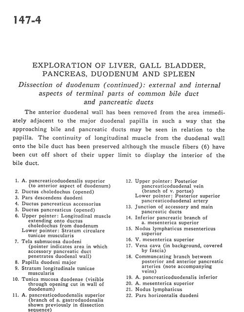
The anterior duodenal wall has been removed from the area immediately adjacent to the major duodenal papilla in such a way that the approaching bile and pancreatic ducts may be seen in relation to the papilla. The continuity of longitudinal muscle from the duodenal wall onto the bile duct has been preserved although the muscle fibres (6) have been cut off short of their upper limit to display the interior of the bile duct.
- Superior pancreaticoduodenal artery (to anterior aspect of duodenum)
- Common bile duct (opened)
- Descending part of duodenum
- Accessory pancreatic duct
- Pancreatic duct (opened)
- Upper pointer: Longitudinal muscle extending onto common bile duct from duodenum Lower pointer: Circular fibers of muscular layer
- Submucosa of duodenum (pointer indicates area in which accessory pancreatic duct penetrates duodenal wall)
- Major duodenal papilla
- Longitudinal muscle layer of stomach
- Muscular layer of duodenum (visible through opening cut in wall of duodenum)
- Superior pancreaticoduodenal artery (branch of gastroduodenal artery shown previously in dissection sequence)
- Upper pointer: Posterior pancreaticoduodenal vein (branch of portal vein) Lower pointer: Posterior superior pancreaticoduodenal artery
- Junction of accessory and main pancreatic ducts
- Inferior pancreatic branch of superior mesenteric artery
- Superior mesenteric lymph node
- Superior mesenteric vein
- Vena cava (in background, covered by fascia)
- Communicating branch between posterior and anterior pancreatic arteries (note accompanying veins)
- Inferior pancreaticoduodenal artery
- Superior mesenteric artery
- Lymph node
- Horizontal part of duodenum