Exploration of liver, gall bladder, pancreas, duodenum and spleen
Interior of gall bladder, bile ducts and pancreatic duct
Stanford holds the copyright to the David L. Bassett anatomical images and has assigned
Creative Commons license Attribution-Share
Alike 4.0 International to all of the images.
For additional information regarding use and permissions,
please contact the Medical History Center.
Image #146-6
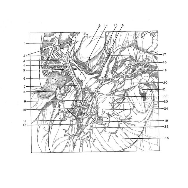
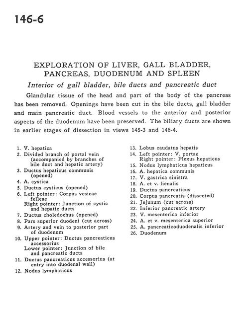
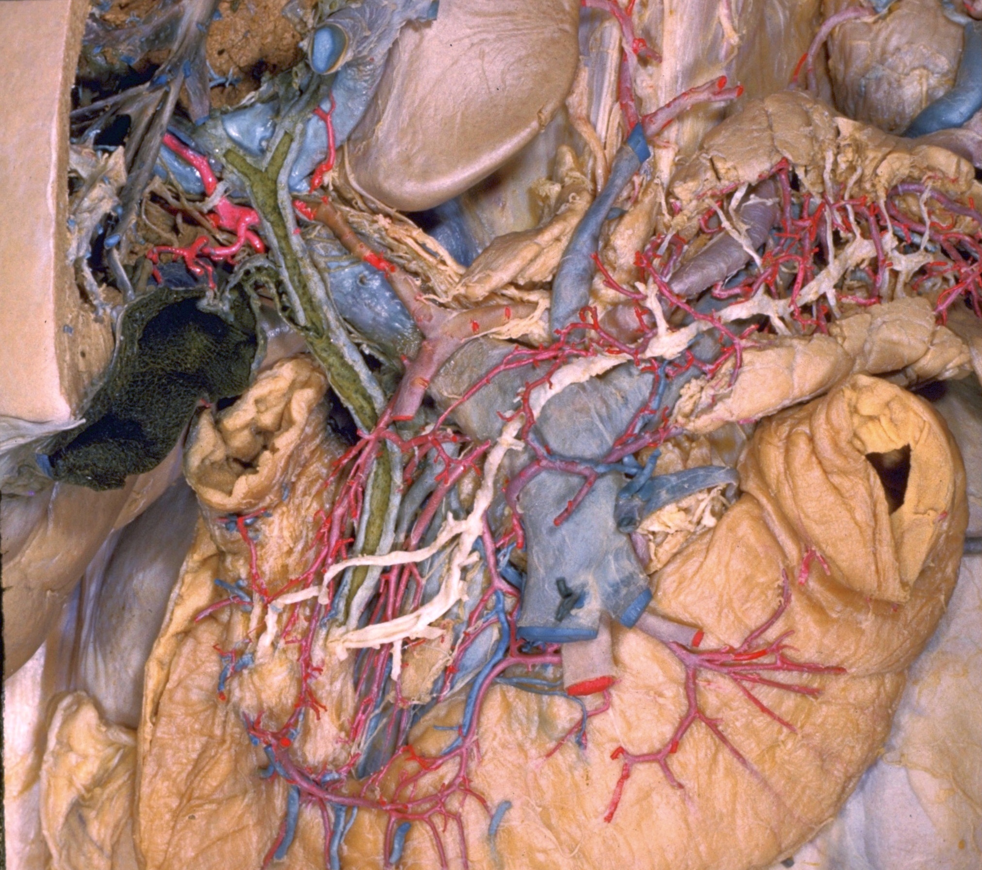
Exploration of liver, gall bladder, pancreas, duodenum and spleen
Interior of gall bladder, bile ducts and pancreatic duct
Glandular tissue of the head and part of the body of the pancreas has been removed. Openings have been cut in the bile ducts, gall bladder and main pancreatic duct. Blood vessels to the anterior and posterior aspects of the duodenum have been preserved. The biliary ducts are shown in earlier stages of dissection in views 145-3 and 146-4
- Hepatic vein
- Divided branch of portal vein (accompanied by branches of bile duct and hepatic artery)
- Common hepatic duct (opened)
- Cystic artery
- Cystic duct (opened)
- Left pointer: Body of gallbladder Right pointer: Junction of cystic and hepatic ducts
- Common bile duct (opened)
- Superior part of duodenum (cut across)
- Artery and vein to posterior part of duodenum
- Upper pointer: Accessory pancreatic duct Lower pointer: Junction of bile and pancreatic ducts
- Accessory pancreatic duct (at entry into duodenal wall)
- Lymph node
- Caudate lobe of liver
- Left pointer: Portal vein Right pointer: Hepatic plexus
- Hepatic lymph node
- Common hepatic artery
- Left gastric vein
- Splenic artery and vein
- Pancreatic duct
- Body of pancreas (dissected)
- Jejunum (cut across)
- Inferior pancreatic artery
- Inferior mesenteric vein
- Superior mesenteric artery and vein
- Inferior pancreaticoduodenal artery
- Duodenum