Exploration of liver, gall bladder, pancreas, duodenum and spleen
Dissection of porta hepatis; cystic duct and cystic artery
Stanford holds the copyright to the David L. Bassett anatomical images and has assigned
Creative Commons license Attribution-Share
Alike 4.0 International to all of the images.
For additional information regarding use and permissions,
please contact the Medical History Center.
Image #145-3
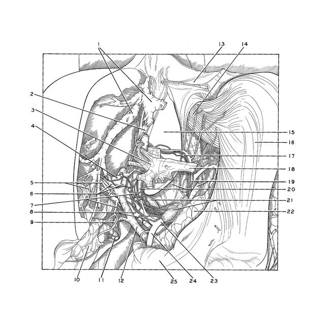
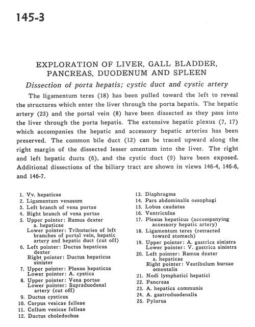
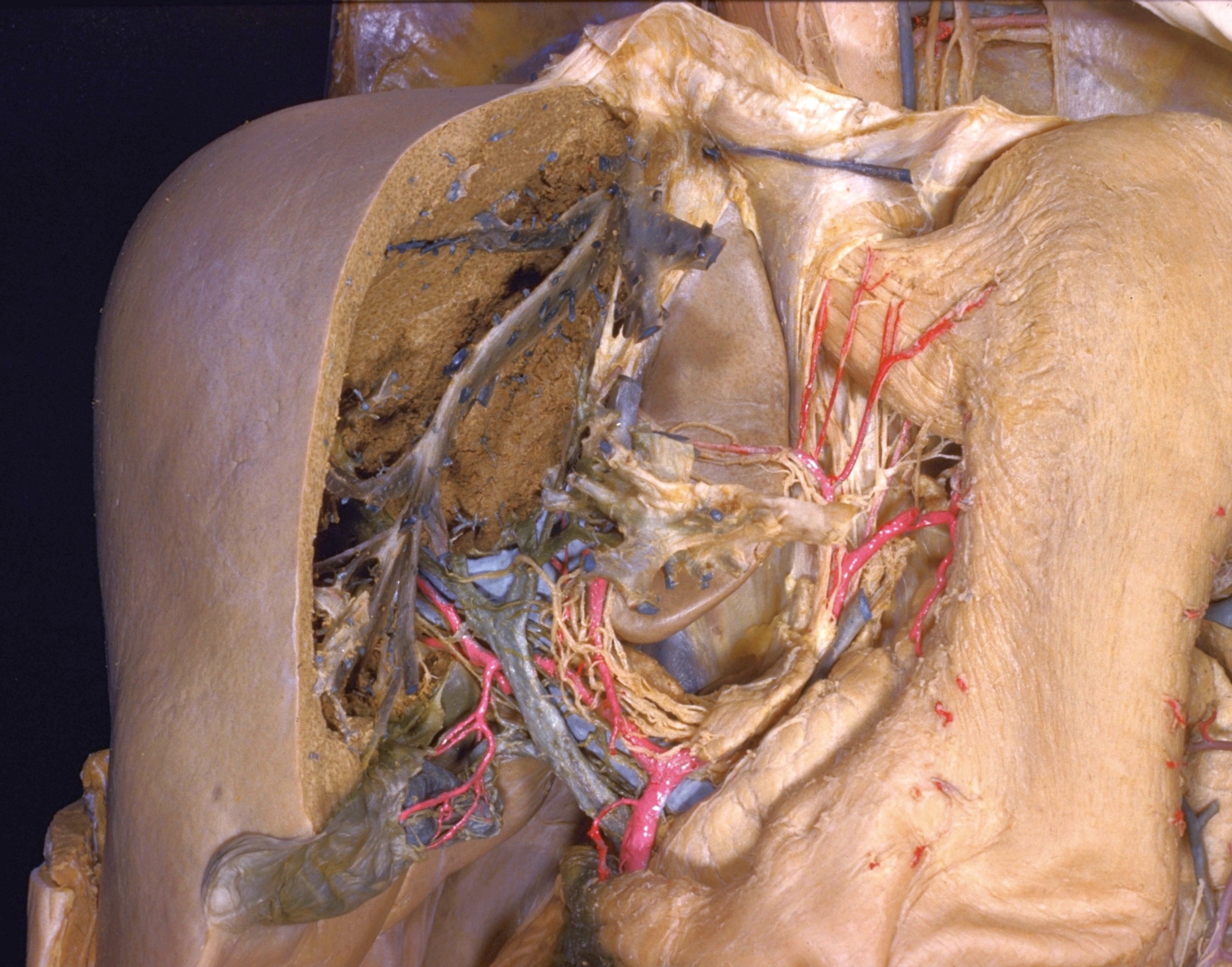
Exploration of liver, gall bladder, pancreas, duodenum and spleen
Dissection of porta hepatis; cystic duct and cystic artery
The ligamentum teres (18) has been pulled toward the left to reveal the structures which enter the liver through the porta hepatis. The hepatic artery (23) and the portal vein (8) have been dissected as they pass into the liver through the porta hepatis. The extensive hepatic plexus (7, 17) which accompanies the hepatic and accessory hepatic arteries has been preserved. The common bile duct (12) can be traced upward along the right margin of the dissected lesser omentum into the liver. The right and left hepatic ducts (6), and the cystic duct (9) have been exposed. Additional dissections of the biliary tract are shown in views 146-4, 146-6 and 146-7.
- Hepatic veins
- Ligamentum venosum
- Left branch of portal vein
- Right branch of portal vein
- Upper pointer: Right branch of hepatic artery Lower pointer: Tributaries of left branches of portal vein, hepatic artery and hepatic duct (cut off)
- Left pointer: Right hepatic duct Right pointer: Left hepatic duct
- Upper pointer: Hepatic plexus Lower pointer: Cystic artery
- Upper pointer: Portal vein Lower pointer: Supraduodenal artery (cut off)
- Cystic duct
- Body of gallbladder
- Neck of gallbladder
- Common bile duct
- Diaphragm
- Abdominal part of esophagus
- Caudate lobe
- Stomach
- Hepatic plexus (accompanying accessory hepatic artery)
- Ligamentum teres (retracted toward stomach)
- Upper pointer: Left gastric artery Lower pointer: Left gastric vein
- Left pointer: Right branch of hepatic artery Right pointer: Vestibule of omental bursa
- Hepatic lymph nodes
- Pancreas
- Common hepatic artery
- Gastroduodenal artery
- Pylorus