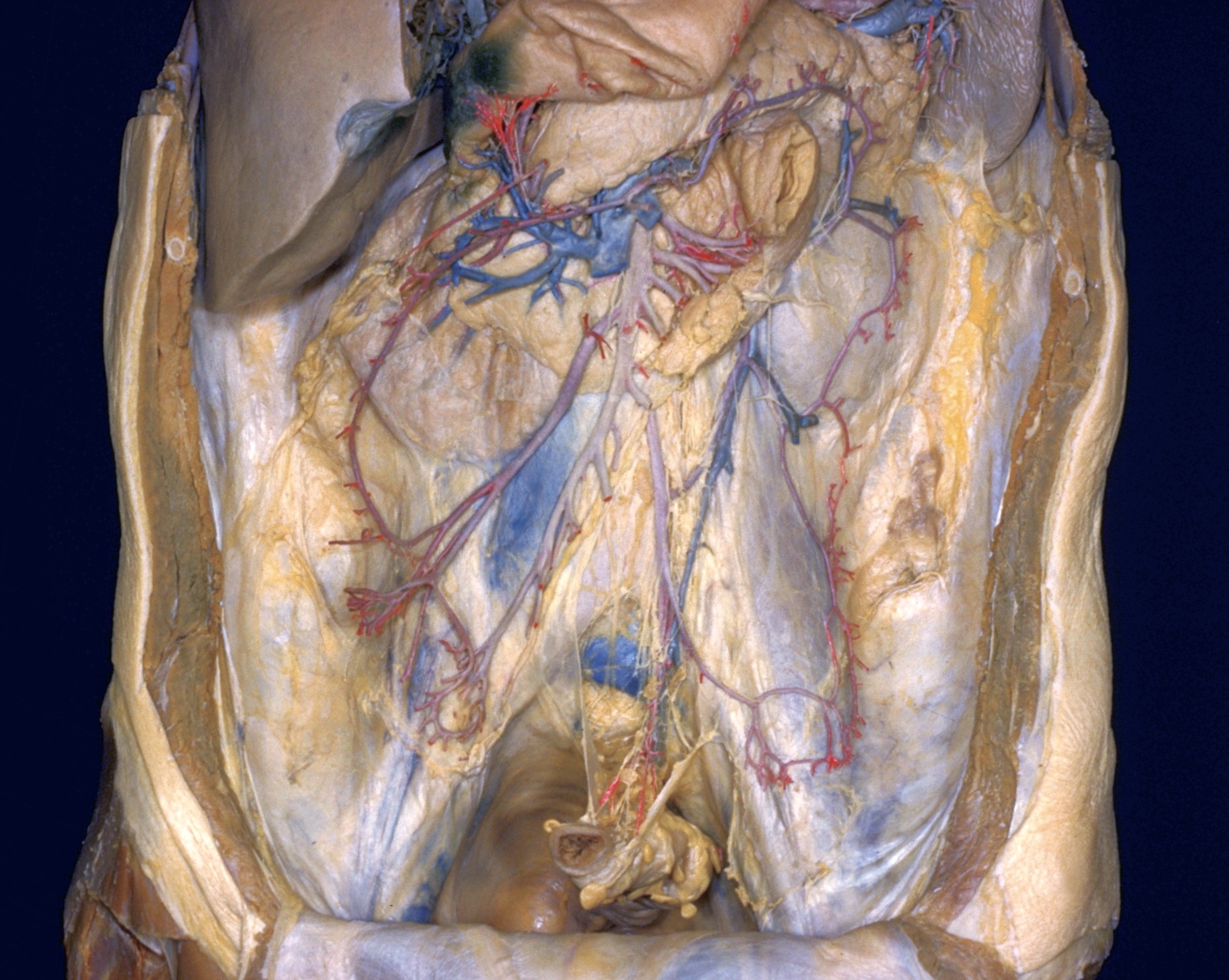
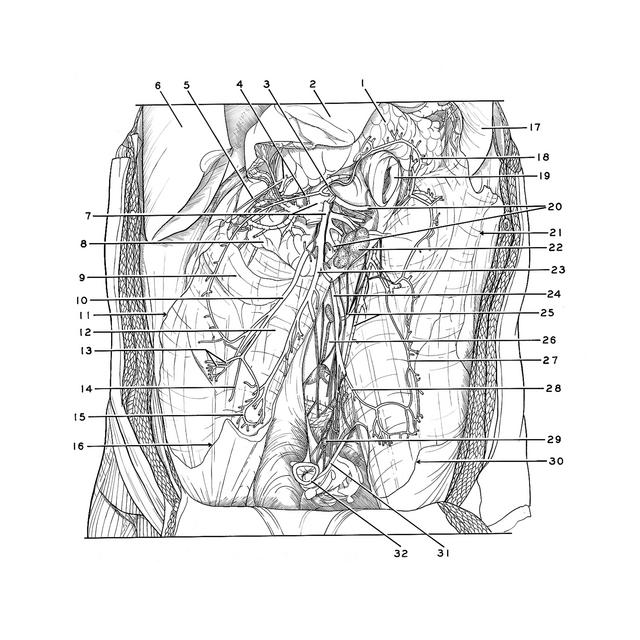
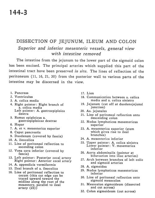
Dissection of jejunum, ileum and colon
Superior and inferior mesenteric vessels, general view with intestine removed
Stanford holds the copyright to the David L. Bassett anatomical images and has assigned
Creative Commons license Attribution-Share
Alike 4.0 International to all of the images.
For additional information regarding use and permissions,
please contact the Medical History Center.
Image #144-3
Dissection of jejunum, ileum and colon
Superior and inferior mesenteric vessels, general view with intestine removed
The intestine from the jejunum to the lower part of the sigmoid colon has been excised. The principal arteries which supplied this part of the intestinal tract have been preserved in situ. The lines of reflection of the peritoneum (11, 16, 21, 30) from the posterior wall to various parts of the intestine may be discerned in the view.
- Pancreas
- Stomach
- Middle colic artery
- Right pointer: Right branch of middle colic artery Left pointer: Right gastroepiploic artery
- Epiploic branch of right gastroepiploic artery
- Liver
- Superior mesenteric artery and vein
- Head of pancreas
- Duodenum (covered by fascia)
- Ileocolic artery
- Line of peritoneal reflection to ascending colon
- Inferior vena cava (covered by fascia)
- Left pointer: Posterior cecal artery Right pointer: Anterior cecal artery
- Appendicular artery
- Ileal branch of ileocolic artery
- Line of peritoneal reflection to cecum (this cut edge can be traced upward toward the midline along the root of the mesentery, parallel to ileal artery (23))
- Spleen
- Communication between middle colic artery and left colic artery
- Jejunum (cut off at duodenojejunal junction)
- Jejunal arteries
- Line of peritoneal reflection onto descending colon
- Superior mesenteric lymph node
- Superior mesenteric artery (stem which gives rise to ileal branches)
- Inferior mesenteric artery
- Upper pointer: Left colic artery Lower pointer: Inferior mesenteric vein
- Abdominal aorta (pointer at bifurcation into iliac arteries)
- Arch between branches of left colic and sigmoid arteries
- Sigmoid artery
- Inferior mesenteric lymph node
- Line of peritoneal reflection onto sigmoid mesocolon
- Mesosigmoid colon (dissected and cut across)
- Sigmoid colon (cut across)