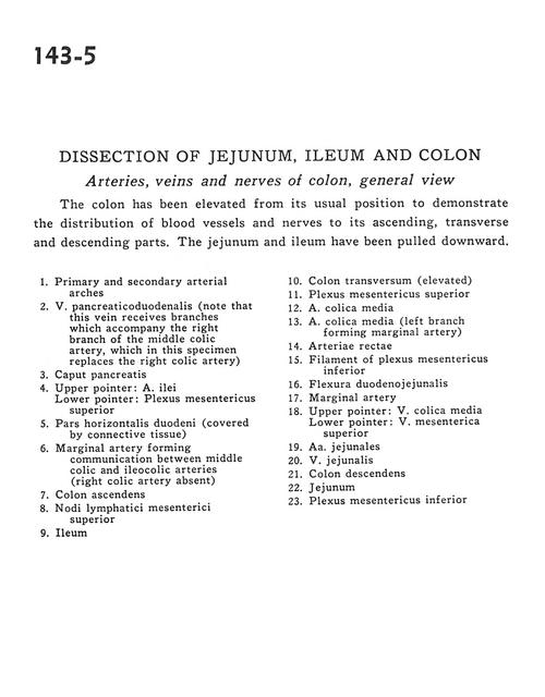
Dissection of jejunum, ileum and colon
Arteries, veins and nerves of colon, general view
Stanford holds the copyright to the David L. Bassett anatomical images and has assigned
Creative Commons license Attribution-Share
Alike 4.0 International to all of the images.
For additional information regarding use and permissions,
please contact the Medical History Center.
Image #143-5
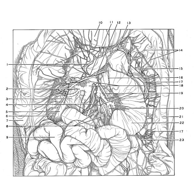
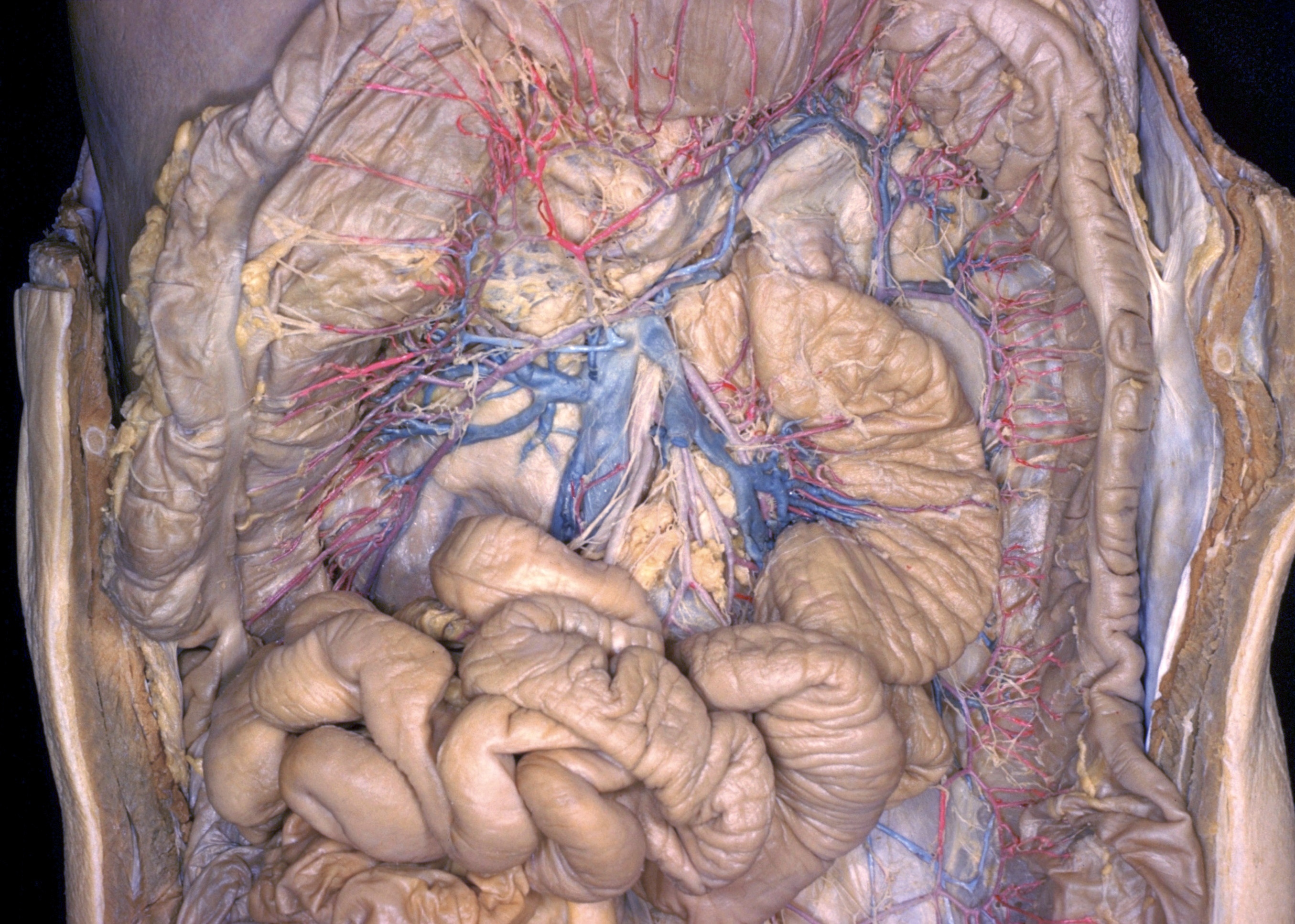
Dissection of jejunum, ileum and colon
Arteries, veins and nerves of colon, general view
The colon has been elevated to form its usual position to demonstrate the distribution of blood vessels and nerves to its ascending, transverse and descending parts. The jejunum and ileum have been pulled downward.
- Primary and secondary arterial arches
- Pancreaticoduodenal vein (note that this vein receives branches which accompany the right branch of the middle colic artery, which in this specimen replaces the right colic artery)
- Head of pancreas
- Upper pointer: Ileal artery Lower pointer: Superior mesenteric plexus
- Horizontal part of duodenum (covered by connective tissue)
- Marginal artery (of Drummond) forming communication between middle colic and ileocolic arteries (right colic artery absent)
- Ascending colon
- Superior mesenteric lymph nodes
- Ileum
- Transverse colon (elevated)
- Superior mesenteric plexus
- Middle colic artery
- Middle colic artery (left branch forming marginal artery)
- Straight arteries
- Filament of inferior mesenteric plexus
- Duodenojejunal flexure
- Marginal artery
- Upper pointer: Middle colic vein Lower pointer: Superior mesenteric vein
- Jejunal arteries
- Jejunal vein
- Descending colon
- Jejunum
- Inferior mesenteric plexus