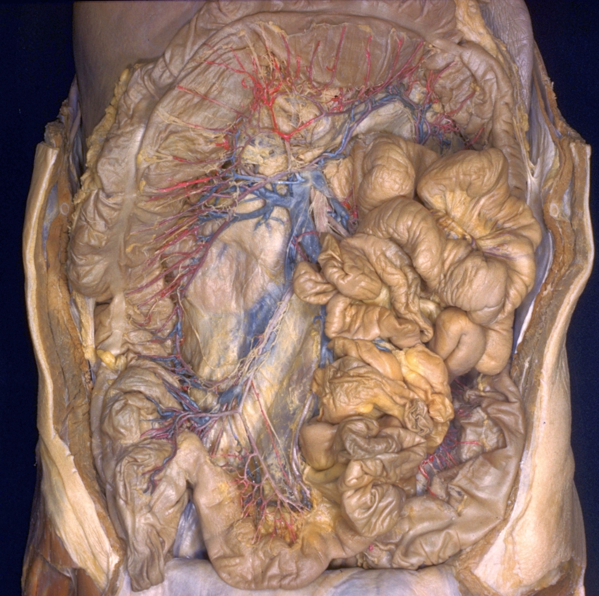
Dissection of jejunum, ileum and colon
Arteries, veins and nerves of ileum, cecum, ascending colon and transverse colon, general view
Stanford holds the copyright to the David L. Bassett anatomical images and has assigned
Creative Commons license Attribution-Share
Alike 4.0 International to all of the images.
For additional information regarding use and permissions,
please contact the Medical History Center.
Image #143-1
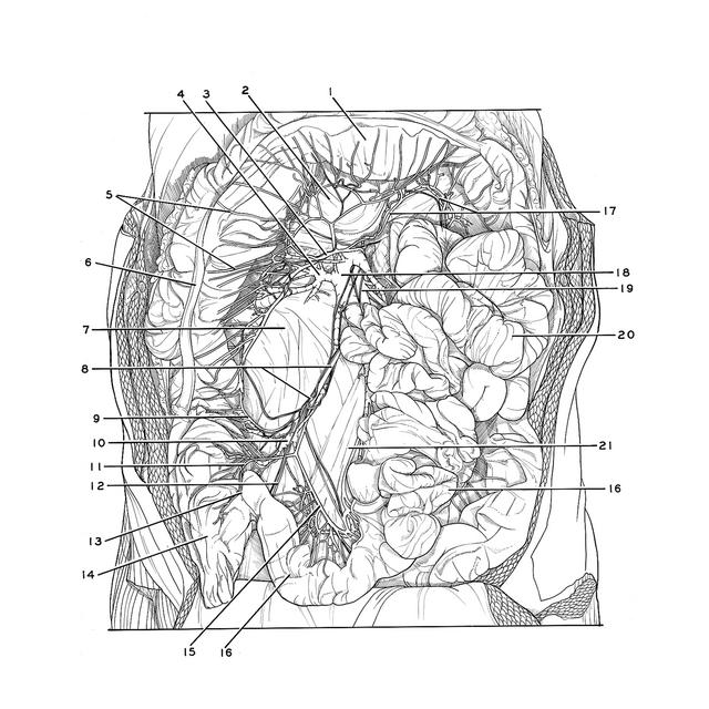
Dissection of jejunum, ileum and colon
Arteries, veins and nerves of ileum, cecum, ascending colon and transverse colon, general view
The jejunum and ileum have been pulled toward the left of the specimen. The transverse colon has been elevated. Lymph nodes and vessels, which were shown in view 141-1, have been removed. The nerves which accompany branches of the superior mesenteric vessels have been preserved insofar as possible.
- Transverse colon (elevated)
- Mesotransverse colon (dissected)
- Middle colic artery (right branch; note absence of a separate right colic artery in this preparation)
- Pancreaticoduodenal vein
- Straight arteries
- Ascending colon (pointer on taenia coli)
- Position of duodenum (covered by fascia)
- Upper pointer: Ileocolic artery Lower pointer: Superior mesenteric plexus
- Colic branch of ileocolic artery
- Ileocolic vein
- Anterior cecal branch of ileocolic artery
- Appendicular artery
- Ileocecal junction
- Cecum
- Ileal branch of ileocolic artery
- Ileum
- Left branch of middle colic artery (accompanied by middle colic vein)
- Superior mesenteric vein
- Superior mesenteric artery (accompanied by superior mesenteric plexus)
- Jejunum
- Mesenteries (dissected)