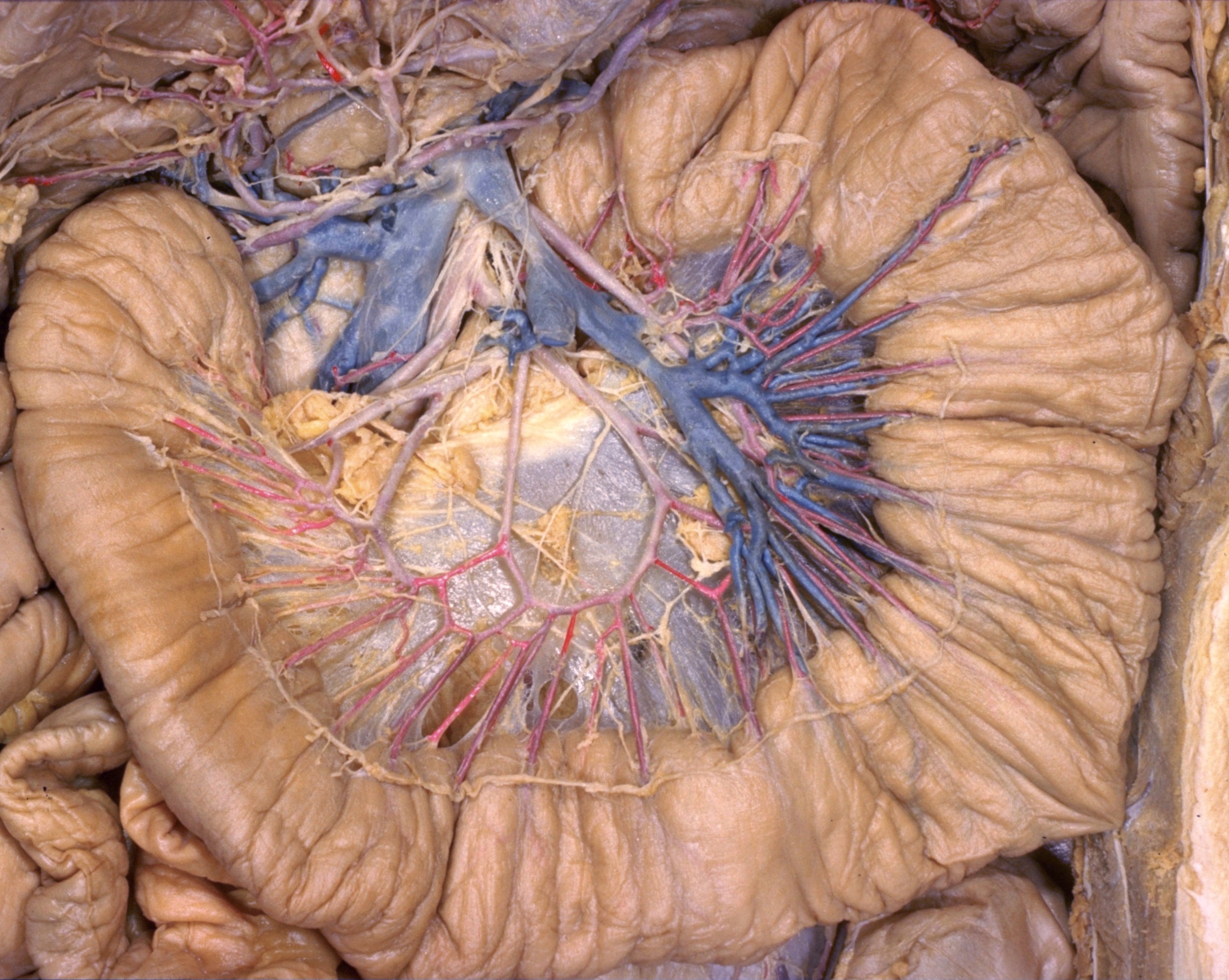
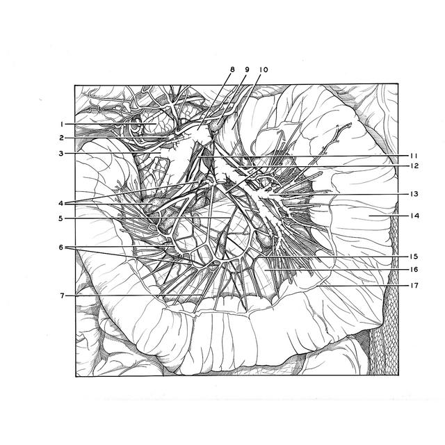
Dissection of jejunum, ileum and colon
Arteries, veins and nerves of upper part of jejunum
Stanford holds the copyright to the David L. Bassett anatomical images and has assigned
Creative Commons license Attribution-Share
Alike 4.0 International to all of the images.
For additional information regarding use and permissions,
please contact the Medical History Center.
Image #142-7
Dissection of jejunum, ileum and colon
Arteries, veins and nerves of upper part of jejunum
The first part of the jejunum has been arranged in a loop with its mesentery dissected to illustrate the manner in which vessels and nerves approach the intestinal wall. The peritoneal layer which formed the left side of the mesentery has been preserved behind the dissected vessels. Lymphatic structures, which are illustrated in view 141-2, have been mostly removed.
- Head of pancreas
- Middle colic artery
- Pancreaticoduodenal vein
- Mesenteric lymph nodes
- Filament of superior mesenteric plexus
- Arterial arches between branches of jejunal arteries (upper pointer, primary arch; lower pointer, secondary arch)
- Straight artery
- Superior mesenteric vein
- Mesenteric lymph node
- Ascending part of duodenum
- Superior mesenteric artery (accompanied by filaments of superior mesenteric plexus)
- Jejunal arteries
- Jejunal vein
- Jejunum
- Superior mesenteric plexus (note archlike arrangement of anastomosing nerve strands)
- Mesenteries (peritoneum of left side of mesentery exposed from within)
- Peritoneum (cut at margin of dissected area)