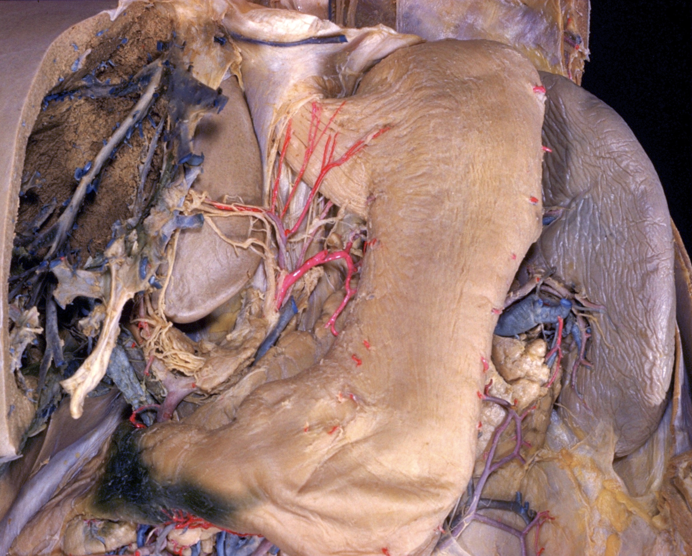
Dissection of stomach
External muscular layer of gastric wall, anterior aspect
Stanford holds the copyright to the David L. Bassett anatomical images and has assigned
Creative Commons license Attribution-Share
Alike 4.0 International to all of the images.
For additional information regarding use and permissions,
please contact the Medical History Center.
Image #142-3

Dissection of stomach
External muscular layer of gastric wall, anterior aspect
The peritoneum has been stripped from the anterior surface of the stomach to display the longitudinally arranged external layer of muscle. This layer is so compact in the region of the pyloric antrum, pyloric canal and pylorus that the direction of its component fibres cannot be easily discerned. Elsewhere, the muscle is formed into coarser fascicles which are clearly visible. The short, cut ends of arteries which protrude from the gastric wall may be identified by reference to the two preceding views.
- Diaphragm
- Cardiac notch
- Margin of esophageal hiatus
- Abdominal part of esophagus
- Esophageal branch of left gastric artery
- Left crus of diaphragm (in background)
- Accessory hepatic artery
- Hepatic vein
- Perivascular fibrous capsule (Glisson's capsule, enclosing branches of portal vein, hepatic artery and bile duct)
- Hepatic plexus
- Upper pointer: Left gastric vein (cut off) Lower pointer: Pancreas
- Celiac lymph node
- Upper pointer: Common bile duct Lower pointer: Portal vein
- Proper hepatic artery
- Gastroduodenal artery
- Right pointer: Pyloric canal Left pointer: Pylorus
- Duodenum
- Fundus of stomach
- Cardiac part of stomach
- Spleen
- Major curvature of stomach
- Anterior wall of stomach (note longitudinal muscle layer of stomach)
- Left gastric artery
- Lesser curvature of stomach
- Incisura angularis
- Tail of pancreas
- Antrum pyloricum