Dissection of stomach
Blood vessels and nerves of lesser curvature of stomach, close-up view
Stanford holds the copyright to the David L. Bassett anatomical images and has assigned
Creative Commons license Attribution-Share
Alike 4.0 International to all of the images.
For additional information regarding use and permissions,
please contact the Medical History Center.
Image #142-1
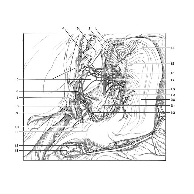
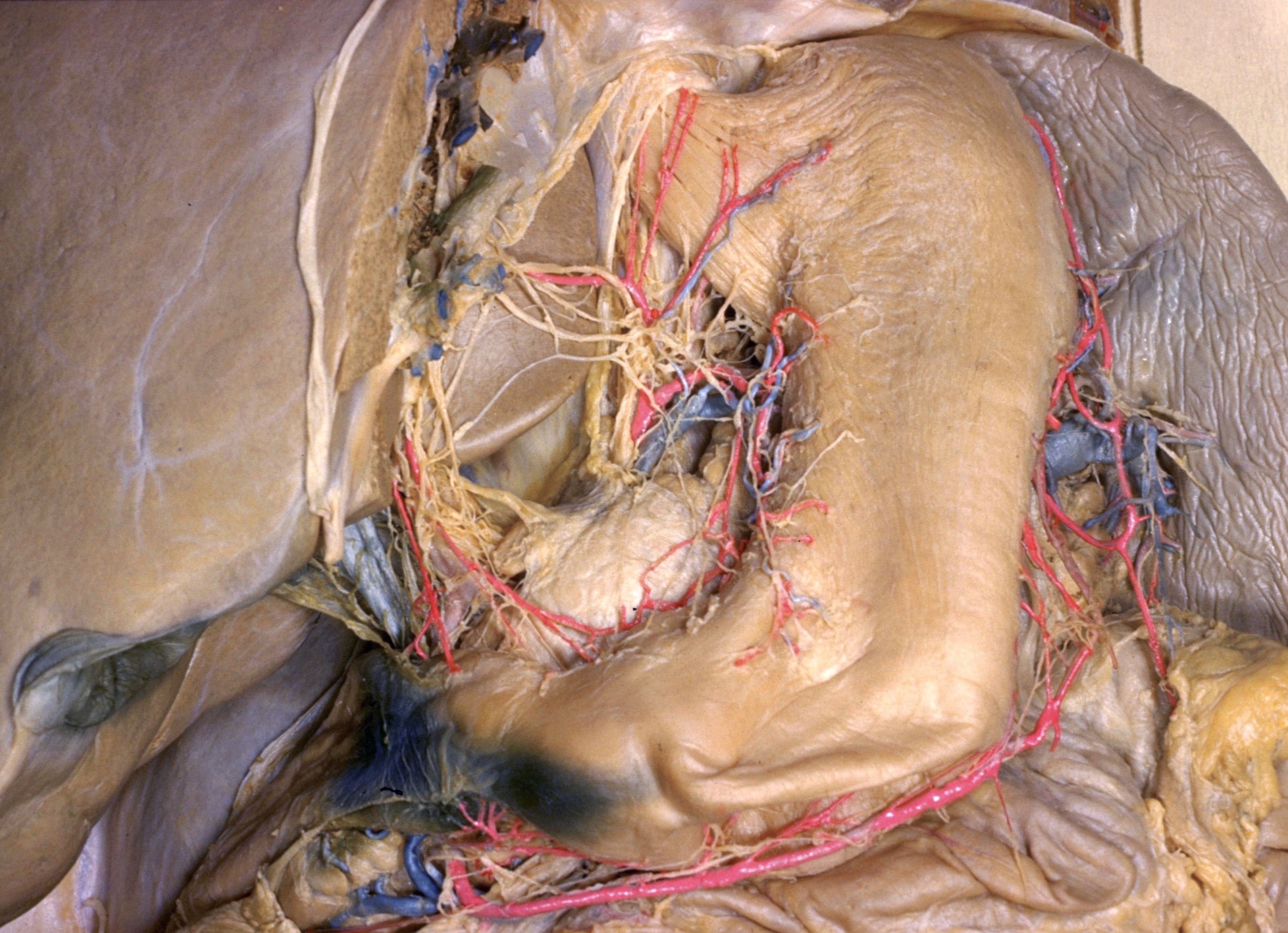
Dissection of stomach
Blood vessels and nerves of lesser curvature of stomach, close-up view
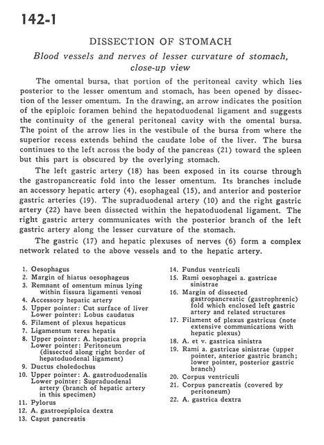
The omental bursa, that portion of the peritoneal cavity which lies posterior to the lesser omentum and stomach, has been opened by dissection of the lesser omentum. In the drawing, an arrow indicates the position of the epiploic foramen behind the hepatoduodenal ligament and suggests the continuity of the general peritoneal cavity with the omental bursa. The point of the arrow lies in the vestibule of the bursa from where the superior recess extends behind the caudate lobe of the liver. The bursa continues to the left across the body of the pancreas (21) toward the spleen but this part is obscured by the overlying stomach.
- Esophagus
- Margin of esophageal hiatus
- Remnant of lesser omentum lying within fissure of ligamentum venosum
- Accessory hepatic artery
- Upper pointer: Cut surface of liver Lower pointer: Caudate lobe
- Filament of hepatic plexus
- Ligamentum teres (of liver)
- Upper pointer: Proper hepatic artery Lower pointer: Peritoneum (dissected along right border of hepatoduodenal ligament)
- Common bile duct
- Upper pointer: Gastroduodenal artery Lower pointer: Supraduodenal artery (branch of hepatic artery in this specimen)
- Pylorus
- Right gastroepiploic artery
- Head of pancreas
- Fundus of stomach
- Esophageal branches left gastric artery
- Margin of dissected gastropancreatic (gastrophrenic) fold which enclosed left gastric artery and related structures
- Filament of gastric plexus (note extensive communications with hepatic plexus)
- Left gastric artery and vein
- Rami left gastric artery (upper pointer, anterior gastric branch; lower pointer, posterior gastric branch)
- Body of stomach
- Body of pancreas (covered by peritoneum)
- Right gastric artery