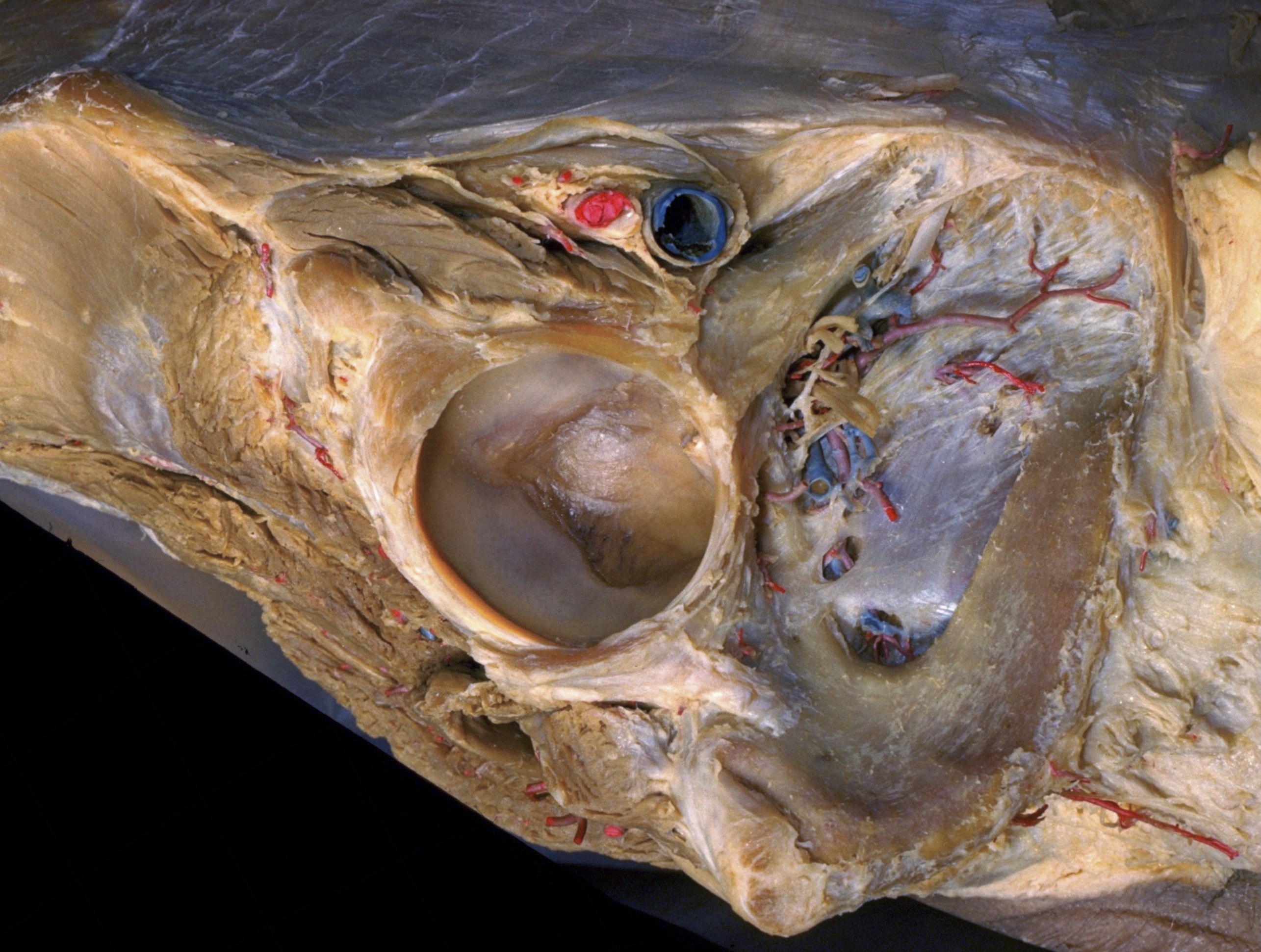
Dissection of female inguinal region
Vascular and muscular compartments deep to inguinal ligament; femoral sheath
Stanford holds the copyright to the David L. Bassett anatomical images and has assigned
Creative Commons license Attribution-Share
Alike 4.0 International to all of the images.
For additional information regarding use and permissions,
please contact the Medical History Center.
Image #138-2
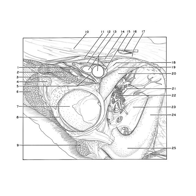
Dissection of female inguinal region
Vascular and muscular compartments deep to inguinal ligament; femoral sheath
The right thigh has been removed by disarticulation at the hip joint. The obturator externus and pectineus muscles have been cut away. Fascia lata (11) has been divided slightly below the inguinal ligament. If this fascia is traced medially in its course toward the pubic tubercle, it is seen to give off a layer which passes deeply to join the medial part of the femoral sheath.
- Iliopectineal arch (iliopectineal ligament)
- Femoral nerve
- Iliopsoas muscle (2 and 3 are the contents of the muscular lacuna)
- Anterior inferior iliac spine (origin of rectus femoris muscle)
- Upper pointer: Femoral sheath (posterior layer, note attachment of this layer to iliopectineal eminence) Lower pointer: Location of iliopectineal eminence
- Gluteus minimus muscle
- Acetabulum
- Labrum of acetabulum
- Obturator internus muscle (cut off)
- Aponeurosis external oblique muscle
- Fascia lata (cut across and elevated)
- Femoral sheath (anterior layer)
- Femoral artery and vein (within venous lacuna)
- Inguinal ligament
- Femoral canal (cut across at its apex canal widens medially within femoral sheath as it approaches level of inguinal ligament)
- Ligamentum teres (of uterus) (emerging through superficial inguinal ring)
- Pubic tubercle
- Location of femoral ring (obscured by medial wall of femoral sheath)
- Pectineal ligament(ligament of Cooper)
- Superior ramus of pubic bone
- Obturator artery and vein
- Obturator nerve
- Obturator membrane
- Inferior pubic ramus
- Ischium