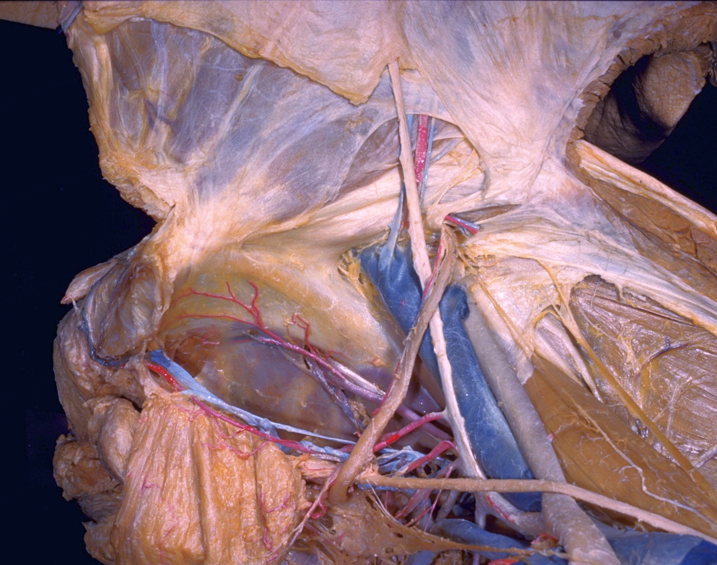
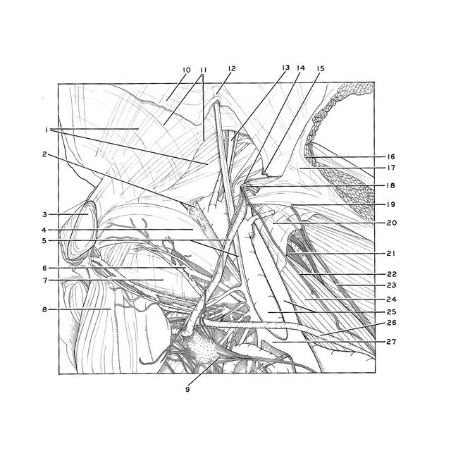
Dissection of male inguinal region and spermatic cord
Internal aspect of deep inguinal ring and related structures of right side
Stanford holds the copyright to the David L. Bassett anatomical images and has assigned
Creative Commons license Attribution-Share
Alike 4.0 International to all of the images.
For additional information regarding use and permissions,
please contact the Medical History Center.
Image #136-6
Dissection of male inguinal region and spermatic cord
Internal aspect of deep inguinal ring and related structures of right side
The specimen is viewed from above and from the left with the anterior abdominal wall pulled forward (toward the top of the photograph). Peritoneum has been preserved in a small area immediately lateral to the deep inguinal ring and, above this, on the interior of the abdominal wall. A small, persistent processus vaginalis (15) is present. It extends as a narrow tube through the deep ingnuinal ring into the upper end of the inguinal canal.
- Right rectus abdominis muscle (upper and lower pointers indicate medial and lateral borders of the muscle)
- Upper pointer: Femoral ring Lower pointer: Lacunar ligament
- Pubic symphysis
- Pectineal ligament (Cooper's ligament)
- Upper pointer: Ductus deferens (accompanied by artery of ductus deferens and filaments from pelvic plexus of nerves) Lower pointer: Lateral umbilical ligament
- Obturator artery (entering obturator canal)
- Fascia covering obturator internus muscles
- Urinary bladder (retracted posteriorly)
- Pelvic plexus
- Peritoneum
- Transversalis fascia
- Medial umbilical fold
- Inferior epigastric artery
- Deep inguinal ring
- Site of opening of patent processus vaginalis
- Transversus abdominis muscle (cut across)
- Transversalis fascia
- Testicular artery (accompanied by testicular veins)
- Area of junction of transversalis fascia and iliac fascia
- Iliac fascia
- Femoral nerve
- Genital branch of genitofemoral nerve
- Femoral branch of genitofemoral nerve (lumboinguinal nerve)
- Psoas major muscle
- External iliac artery and vein
- Ureter
- Internal iliac artery