Dissection of anterolateral abdominal wall
Relation of internal oblique muscle to sheath of rectus at costal margin
Stanford holds the copyright to the David L. Bassett anatomical images and has assigned
Creative Commons license Attribution-Share
Alike 4.0 International to all of the images.
For additional information regarding use and permissions,
please contact the Medical History Center.
Image #133-7
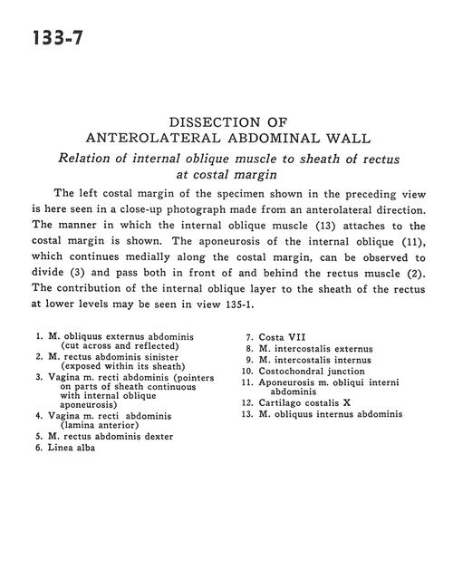
Dissection of anterolateral abdominal wall
Relation of internal oblique muscle to sheath of rectus at costal margin
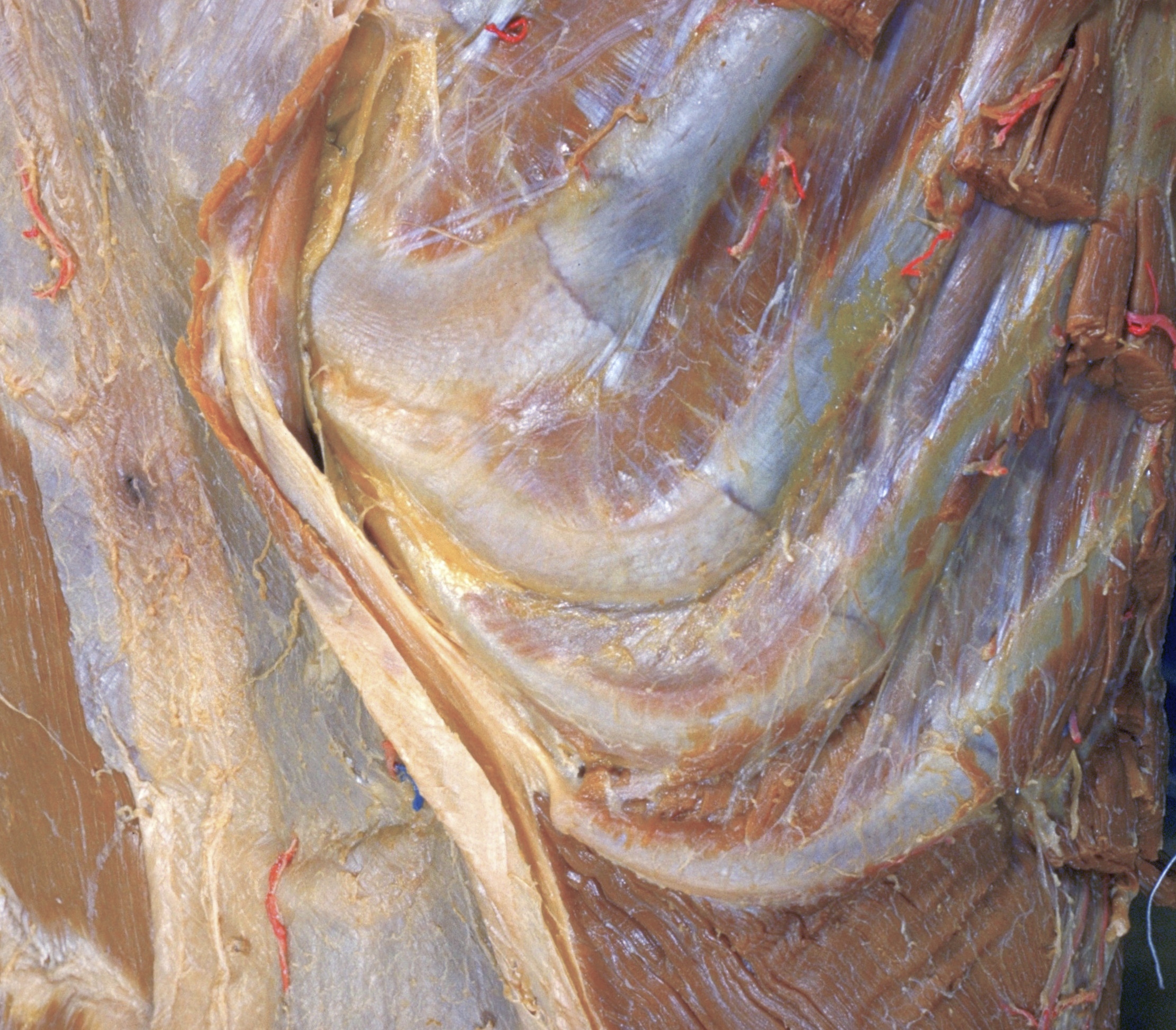
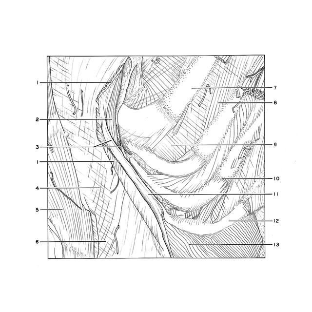
The left costal margin of the specimen shown in the preceding view is here seen in a close-up photograph made from an anterolateral direction. The manner in which the internal oblique muscle (13) attaches to the costal margin is shown. The aponeurosis of the internal oblique (11), which continues medially along the costal margin, can be observed to divide (3) and pass both in front of and behind the rectus muscle (2). The contribution of the internal oblique layer to the sheath of the rectus at lower levels may be seen in view 135-1.
- External oblique muscle (cut across and reflected)
- Left rectus abdominis muscle (exposed within its sheath)
- Sheath of rectus abdominis muscle (pointers on parts of sheath continuous with internal oblique aponeurosis)
- Sheath of rectus abdominis muscle (anterior layer)
- Right rectus abdominis muscle
- Linea alba
- Rib VII
- External intercostal muscle
- Internal intercostal muscle
- Costochondral junction
- Aponeurosis of internal oblique muscle
- Costal cartilage X
- Internal oblique muscle