Dissection of thorax from a posterior approach
Thoracic viscera.
Stanford holds the copyright to the David L. Bassett anatomical images and has assigned
Creative Commons license Attribution-Share
Alike 4.0 International to all of the images.
For additional information regarding use and permissions,
please contact the Medical History Center.
Image #132-6
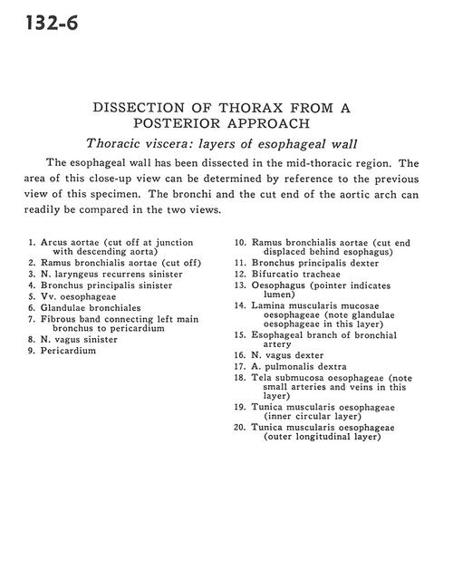
Dissection of thorax from a posterior approach
Thoracic viscera.
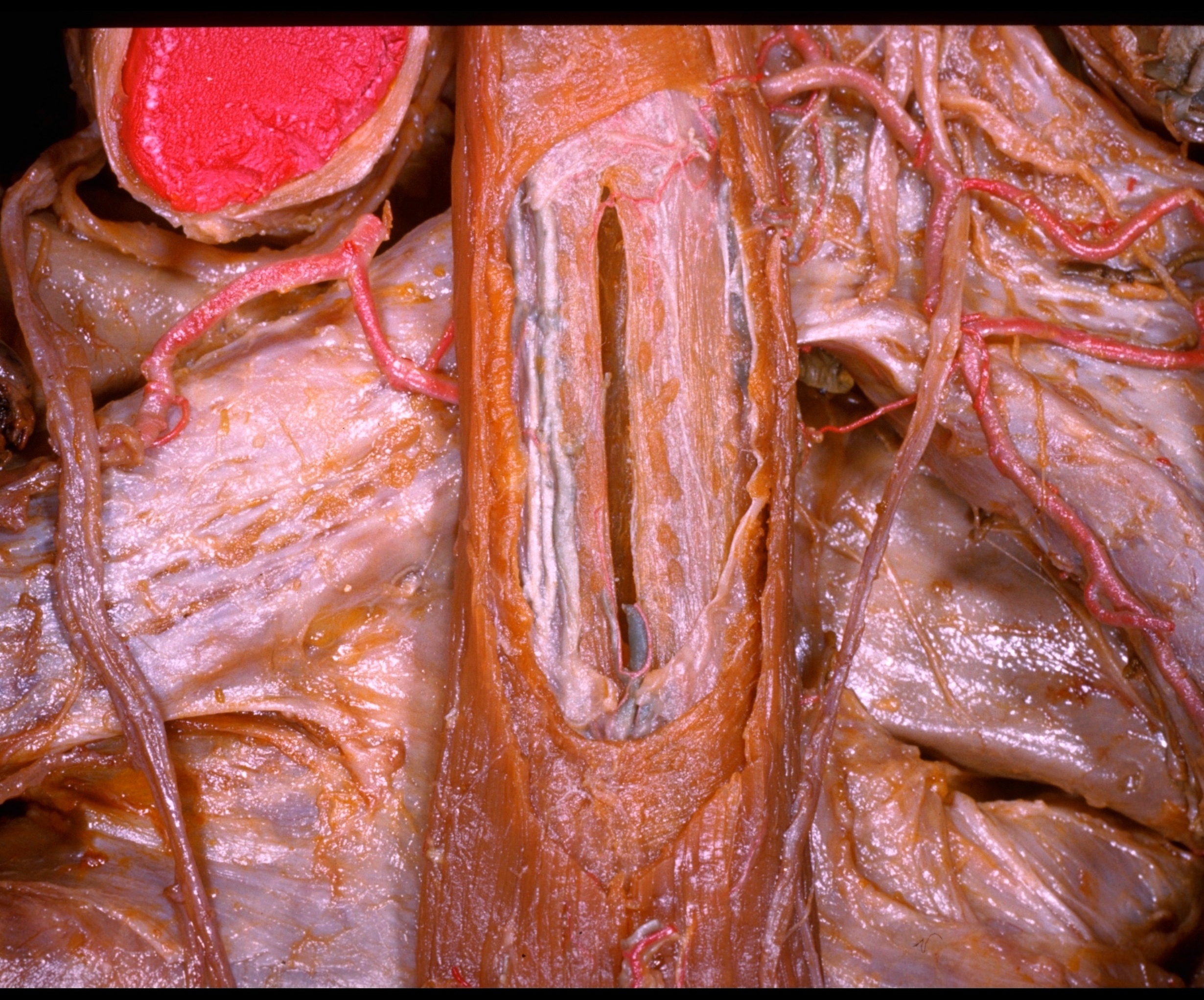
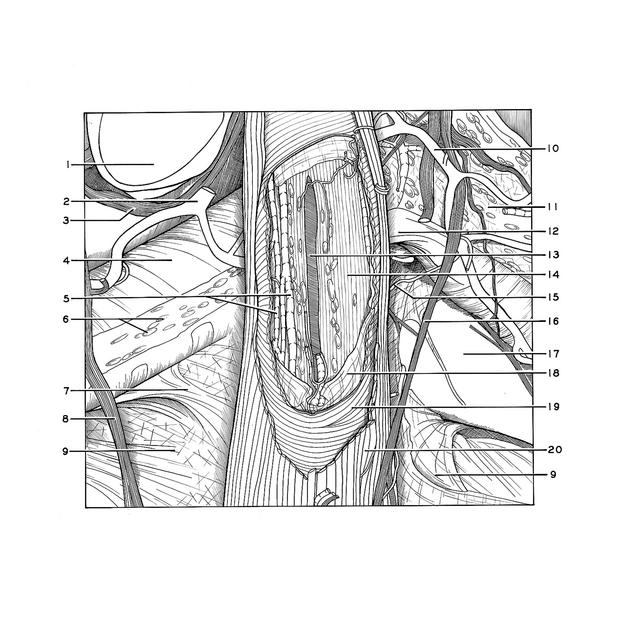
The esophageal wall has been dissected in the mid-thoracic region. The area of this close-up view can be determined by reference to the previous view of this specimen. The bronchi and the cut end of the aortic arch can readily be compared in the two views.
- Aortic arch (cut off at junction with descending aorta)
- Bronchial branch of aorta (cut off)
- Recurrent laryngeal nerve left
- Left main bronchus
- Esophageal veins
- Bronchial glands
- Fibrous band connecting left main bronchus to pericardium
- Vagus nerve left
- Pericardium
- Bronchial branch of aorta (cut end displaced behind esophagus)
- Right main bronchus
- Bifurcation of trachea
- Esophagus (pointer indicates lumen)
- Muscular mucosa of esophagus (note esophageal glands in this layer)
- Esophageal branch of bronchial artery
- Right vagus nerve
- Right pulmonary artery
- Esophageal submucosa (note small arteries and veins in this layer)
- Muscular coat of esophagus (inner circular layer)
- Muscular coat of esophagus (outer longitudinal layer)