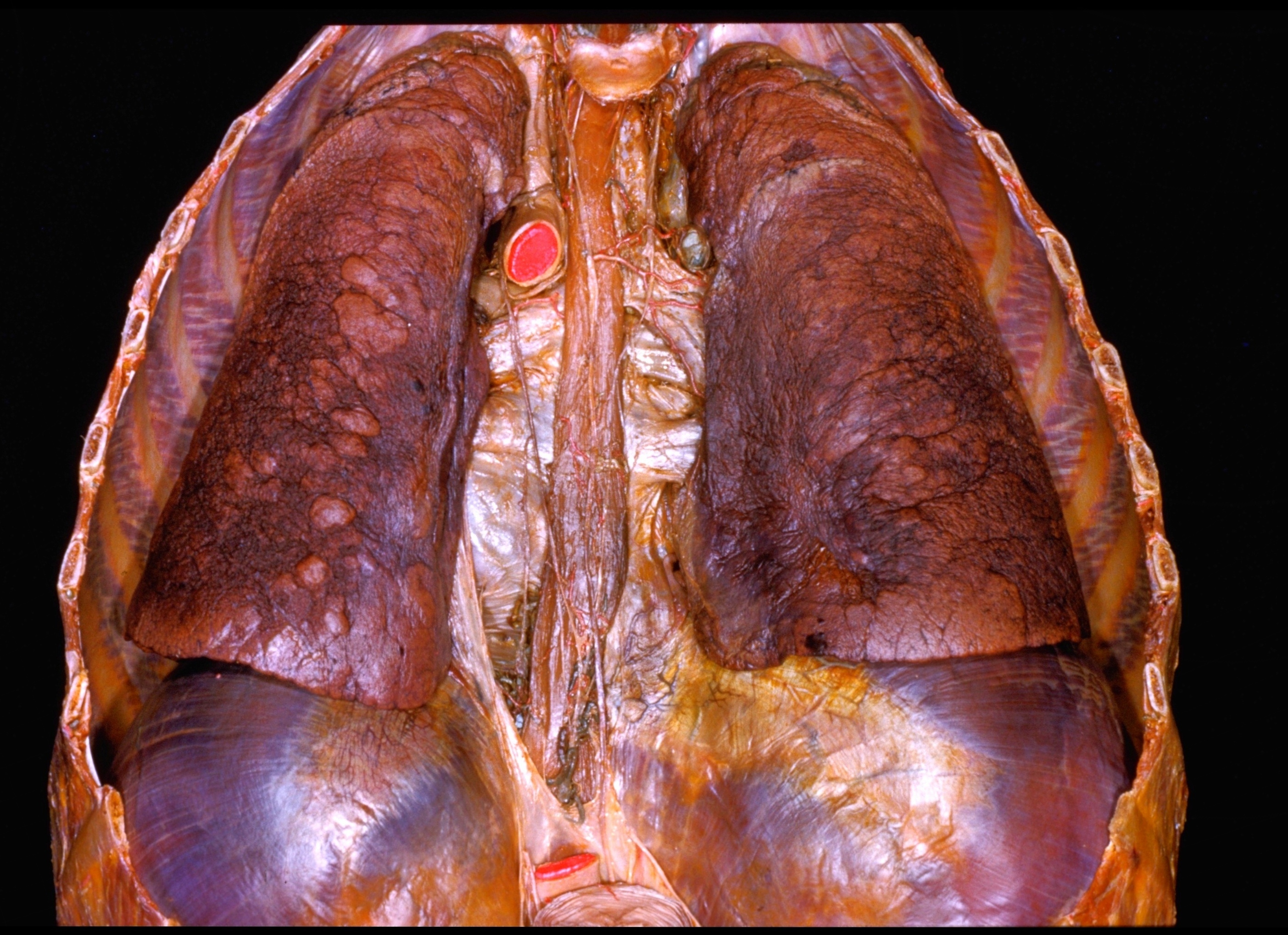
Dissection of thorax from a posterior approach
Thoracic viscera.
Stanford holds the copyright to the David L. Bassett anatomical images and has assigned
Creative Commons license Attribution-Share
Alike 4.0 International to all of the images.
For additional information regarding use and permissions,
please contact the Medical History Center.
Image #132-5
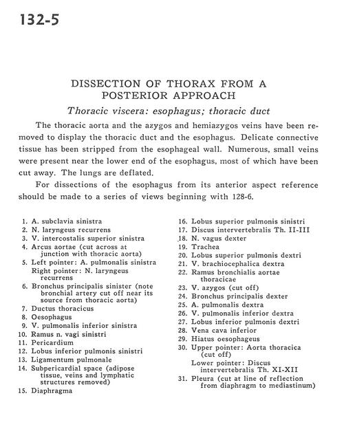
Dissection of thorax from a posterior approach
Thoracic viscera.
The thoracic aorta and the azygos and hemiazygos veins have been removed to display the thoracic duct and the esophagus. Delicate connective tissue has been stripped from the esophageal wall. Numerous, small veins were present near the lower end of the esophagus, most of which have been cut away. The lungs are deflated.
- Left subclavian artery
- Recurrent laryngeal nerve
- Left superior intercostal vein
- Aortic arch (cut across at junction with thoracic aorta)
- Left pointer: Left pulmonary artery Right pointer: Recurrent laryngeal nerve
- Left main bronchus (note bronchial artery cut off near its source from thoracic aorta)
- Thoracic duct
- Esophagus
- Left inferior pulmonary vein
- Vagus nerve left
- Pericardium
- Lower lobe left lung
- Pulmonary ligament
- Subpericardial space (adipose tissue, veins and lymphatic structures removed)
- Diaphragm
- Upper lobe left lung
- Intervertebral disc Th. II-III
- Vagus nerve right
- Trachea
- Upper lobe right lung
- Right brachiocephalic vein
- Bronchial branch of thoracic aorta
- Azygos vein (cut off)
- Right main bronchus
- Right pulmonary artery
- Right inferior pulmonary vein
- Inferior lobe right lung
- Inferior vena cava
- Esophageal hiatus
- Upper pointer: Thoracic aorta (cut off) Lower pointer: Intervertebral disc Th. XI-XII
- Pleura (cut at line of reflection from diaphragm to mediastinum)