Dissection of thorax from a posterior approach
Ligamentous attachment of mediastinal structures to vertebral column
Stanford holds the copyright to the David L. Bassett anatomical images and has assigned
Creative Commons license Attribution-Share
Alike 4.0 International to all of the images.
For additional information regarding use and permissions,
please contact the Medical History Center.
Image #132-3
Dissection of thorax from a posterior approach
Ligamentous attachment of mediastinal structures to vertebral column
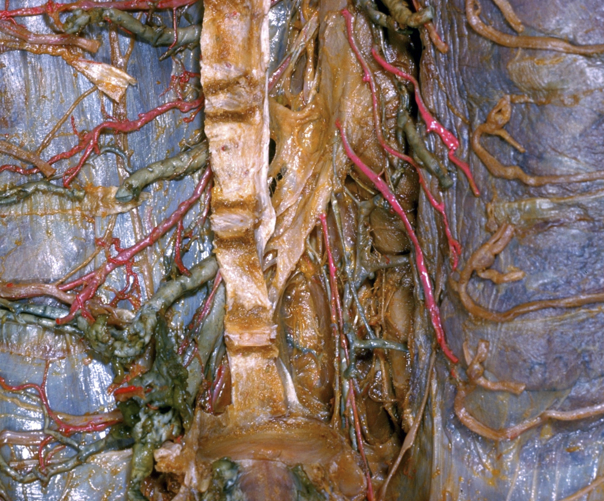
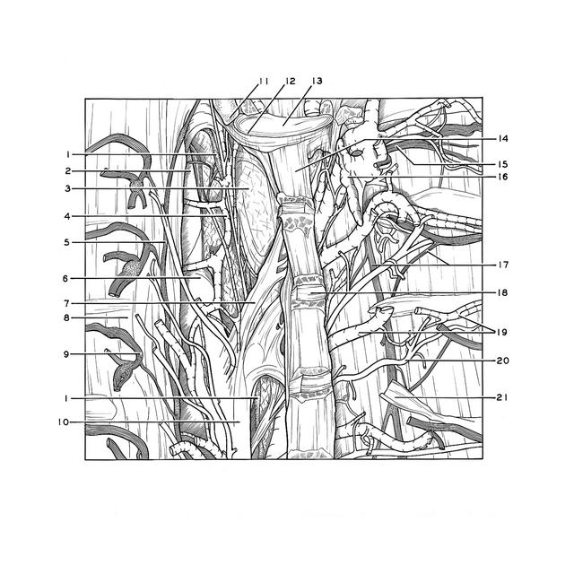
The upper part of the specimen shown in 131-7 is presented in a close-up view with the anterior longitudinal ligament (14) retracted to the right to expose a heavy, ligamentous band which extends down to the aorta from the level of the third and fourth thoracic vertebral bodies. The vertebral attachments of this band blend with the anterior longitudinal ligament near the midline. Inferiorly the ligament blends with the adventitia of the posterior part of the aortic arch and the upper thoracic aorta. It can also be traced forward to where it blends with fascia of the esophagus and trachea, although this arrangement is not visible in this view. This suspensory structure is shown elsewhere in the dissection of another specimen (128-1).
- Thoracic duct
- Left subclavian artery
- Fascia of superior mediastinal space
- Left superior intercostal vein
- Left sympathetic trunk
- Aortic arch
- Ligamentous band (see description above)
- Costal pleura
- Intercostal nerve V
- Thoracic aorta
- Costal facet vertebra Th. II
- Anulus fibrosus
- Nucleus pulposus (12-13 comprise the intervertebral disc)
- Anterior longitudinal ligament
- Intercostal nerve III
- Veins surrounding nerve roots in intervertebral foramen (bones removed)
- Left pointer: Right sympathetic trunk Right pointer: Ramus communicans
- Anulus fibrosus (remnant)
- Posterior intercostal vein V
- Posterior intercostal artery V
- Ligamentum capitis costae radiatum (preserved with periosteum of rib)