Dissection of mediastinum and paravertebral structures
Upper thoracic part of left sympathetic trunk; intercostal vessels; ligamentous band between vertebrae and mediastinal structures
Stanford holds the copyright to the David L. Bassett anatomical images and has assigned
Creative Commons license Attribution-Share
Alike 4.0 International to all of the images.
For additional information regarding use and permissions,
please contact the Medical History Center.
Image #128-1
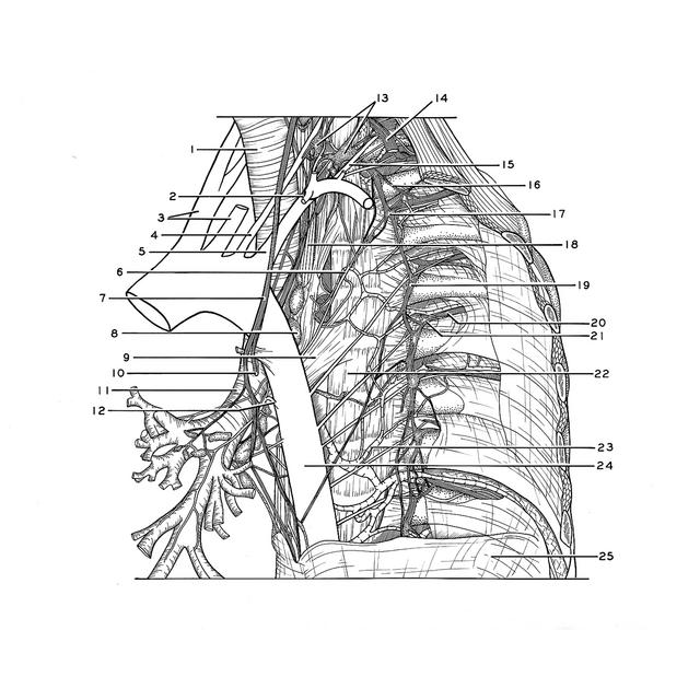
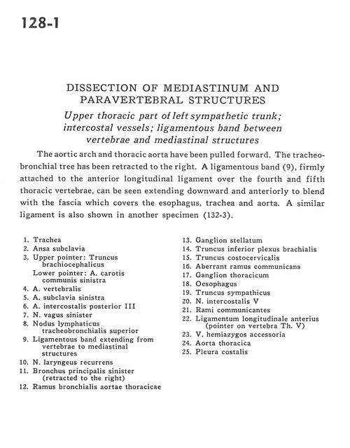
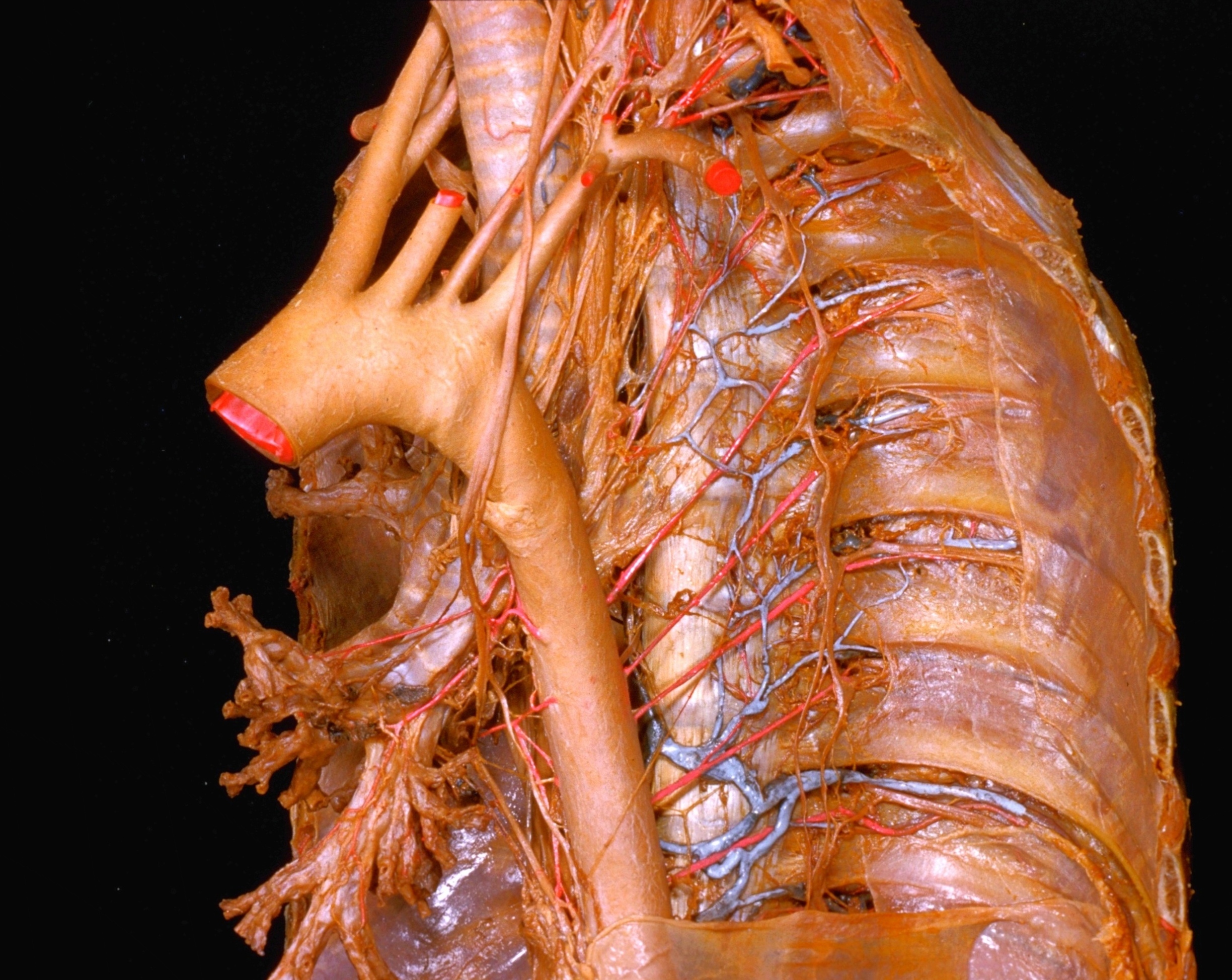
Dissection of mediastinum and paravertebral structures
Upper thoracic part of left sympathetic trunk; intercostal vessels; ligamentous band between vertebrae and mediastinal structures
The aortic arch and thoracic aorta have been pulled forward. The tracheobronchial tree has been retracted to the right. A ligamentous band (9), firmly attached to the anterior longitudinal ligament over the fourth and fifth thoracic vertebrae, can be seen extending downward and anteriorly to blend with the fascia which covers the esophagus, trachea and aorta. A similar ligament is also shown in another specimen (132-3).
- Trachea
- Ansa subclavia
- Upper pointer: Brachiocephalic trunk Lower pointer: Left common carotid artery
- Vertebral artery
- Left subclavian artery
- Posterior intercostal artery III
- Left vagus nerve
- Superior tracheobronchial lymph node
- Ligamentous band extending from vertebrae to mediastinal structures
- Recurrent laryngeal nerve
- Left main bronchus (retracted to the right)
- Bronchial branch of thoracic aorta
- Stellate ganglion
- Inferior trunk brachial plexus
- Costocervical trunk
- Aberrant ramus communicans
- Thoracic ganglion
- Esophagus
- Sympathetic trunk
- Intercostal nerve V
- Rami communicantes
- Anterior longitudinal ligament (pointer on vertebra Th. V)
- Accessory hemiazygos vein
- Thoracic aorta
- Costal pleura