Dissection of lungs in situ
General view of vascular and bronchial distribution within lungs.
Stanford holds the copyright to the David L. Bassett anatomical images and has assigned
Creative Commons license Attribution-Share
Alike 4.0 International to all of the images.
For additional information regarding use and permissions,
please contact the Medical History Center.
Image #123-7
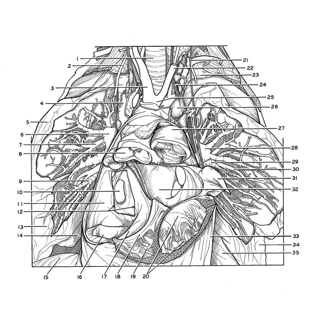
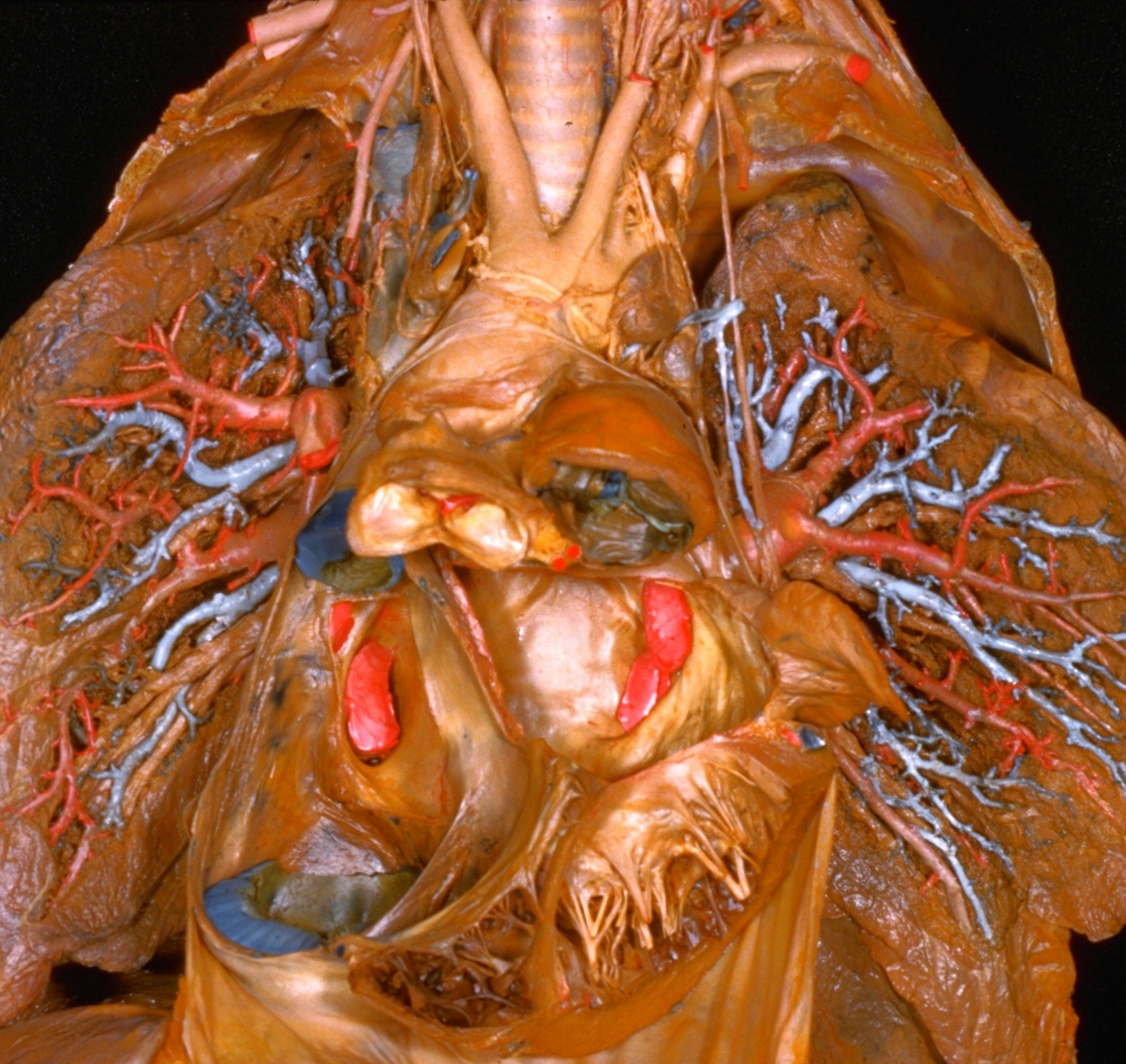
Dissection of lungs in situ
General view of vascular and bronchial distribution within lungs.
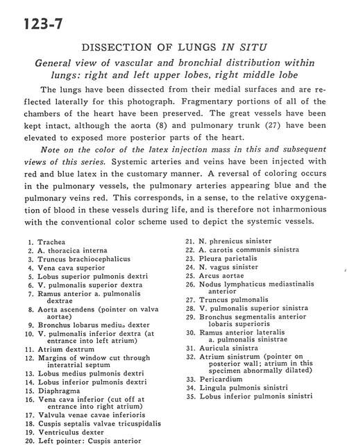
The lungs have been dissected from their medial surfaces and are reflected laterally for this photograph. Fragmentary portions of all of the chambers of the heart have been preserved. The great vessels have been kept intact, although the aorta (8) and pulmonary trunk (27) have been elevated to expose more posterior parts of the heart.
- Trachea
- Internal thoracic artery
- Brachiocephalic trunk
- Superior vena cava
- Upper lobe right lung
- Right superior pulmonary vein
- Ramus anterior right pulmonary artery
- Ascending aorta (pointer on aortic valve)
- Bronchus of right middle lobe
- Right inferior pulmonary vein (at entrance into left atrium)
- Right atrium
- Margins of window cut through interatrial septum
- Middle lobe right lung
- Inferior lobe right lung
- Diaphragm
- Inferior vena cava (cut off at entrance into right atrium)
- Valve of inferior vena cava
- Septal (medial) cusp of tricuspid valve
- Right ventricle
- Left pointer: Anterior cusp of mitral valve Right pointer: Left ventricle
- Left phrenic nerve
- Left common carotid artery
- Parietal pleura
- Left vagus nerve
- Aortic arch
- Anterior mediastinal lymph node
- Pulmonary trunk
- Left superior pulmonary vein
- Anterior segmental bronchus of upper lobe
- Anterior lateral branch left pulmonary artery
- Left auricle
- Left atrium (pointer on posterior wall atrium in this specimen abnormally dilated)
- Pericardium
- Lingula of left lung
- Lower lobe left lung