Detailed dissection of heart
Coronary sinus, external and internal aspects
Stanford holds the copyright to the David L. Bassett anatomical images and has assigned
Creative Commons license Attribution-Share
Alike 4.0 International to all of the images.
For additional information regarding use and permissions,
please contact the Medical History Center.
Image #121-2
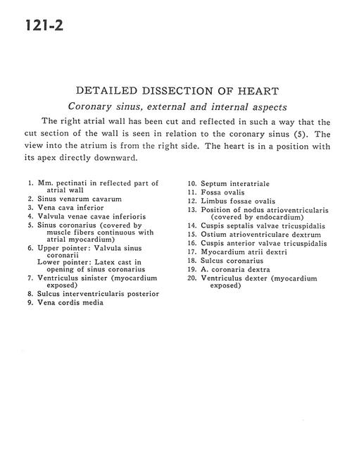
Detailed dissection of heart
Coronary sinus, external and internal aspects
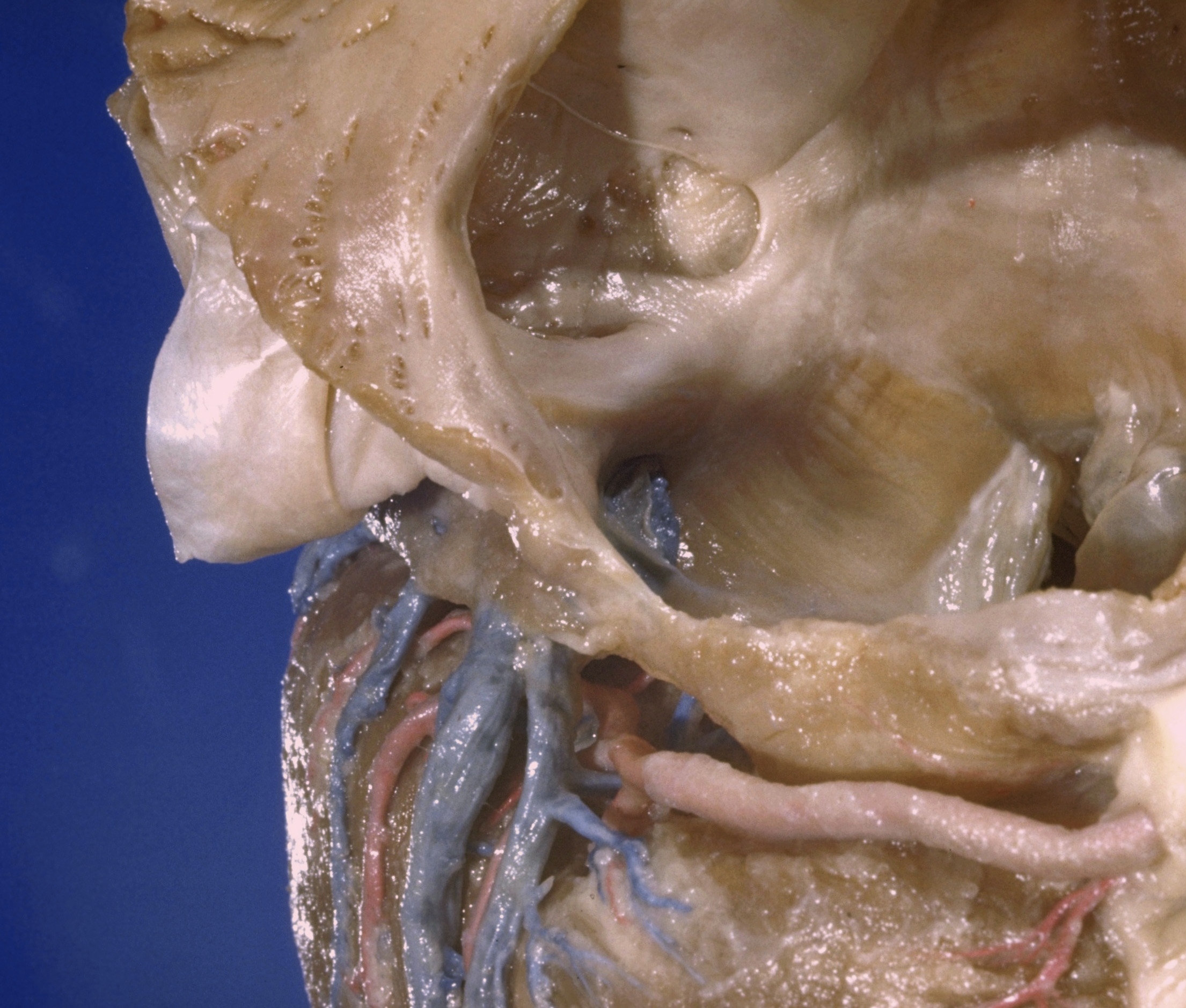
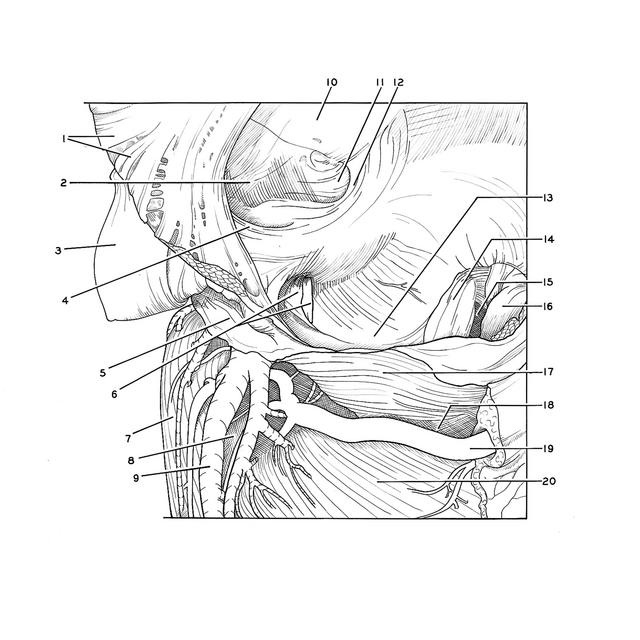
The right atrial wall has been cut and reflected in such a way that the cut section of the wall is seen in relation to the coronary sinus (5). The view into the atrium is from the right side. The heart is in a position with its apex directly downward.
- Pectinate muscles in reflected part of atrial wall
- Sinus of vena cava
- Inferior vena cava
- Valve of inferior vena cava
- Coronary sinus (covered by muscle fibers continuous with atrial myocardium)
- Upper pointer: Valve of coronary sinus Lower pointer: Latex cast in opening of coronary sinus
- Left ventricle (myocardium exposed)
- Posterior interventricular sulcus
- Middle cardiac vein
- Interatrial septum
- Fossa ovalis
- Border of fossa ovalis
- Position of atrioventricular node (covered by endocardium)
- Septal (medial) cusp of tricuspid valve
- Right atrioventricular opening
- Anterior cusp tricuspid valve
- Myocardium right atrium
- Coronary sulcus
- Right coronary artery
- Right ventricle (myocardium exposed)