Exploration of the brain from its superior aspect
Lateral and medial geniculate bodies; hippocampal structures
Stanford holds the copyright to the David L. Bassett anatomical images and has assigned
Creative Commons license Attribution-Share
Alike 4.0 International to all of the images.
For additional information regarding use and permissions,
please contact the Medical History Center.
Image #12-3
Exploration of the brain from its superior aspect
Lateral and medial geniculate bodies; hippocampal structures
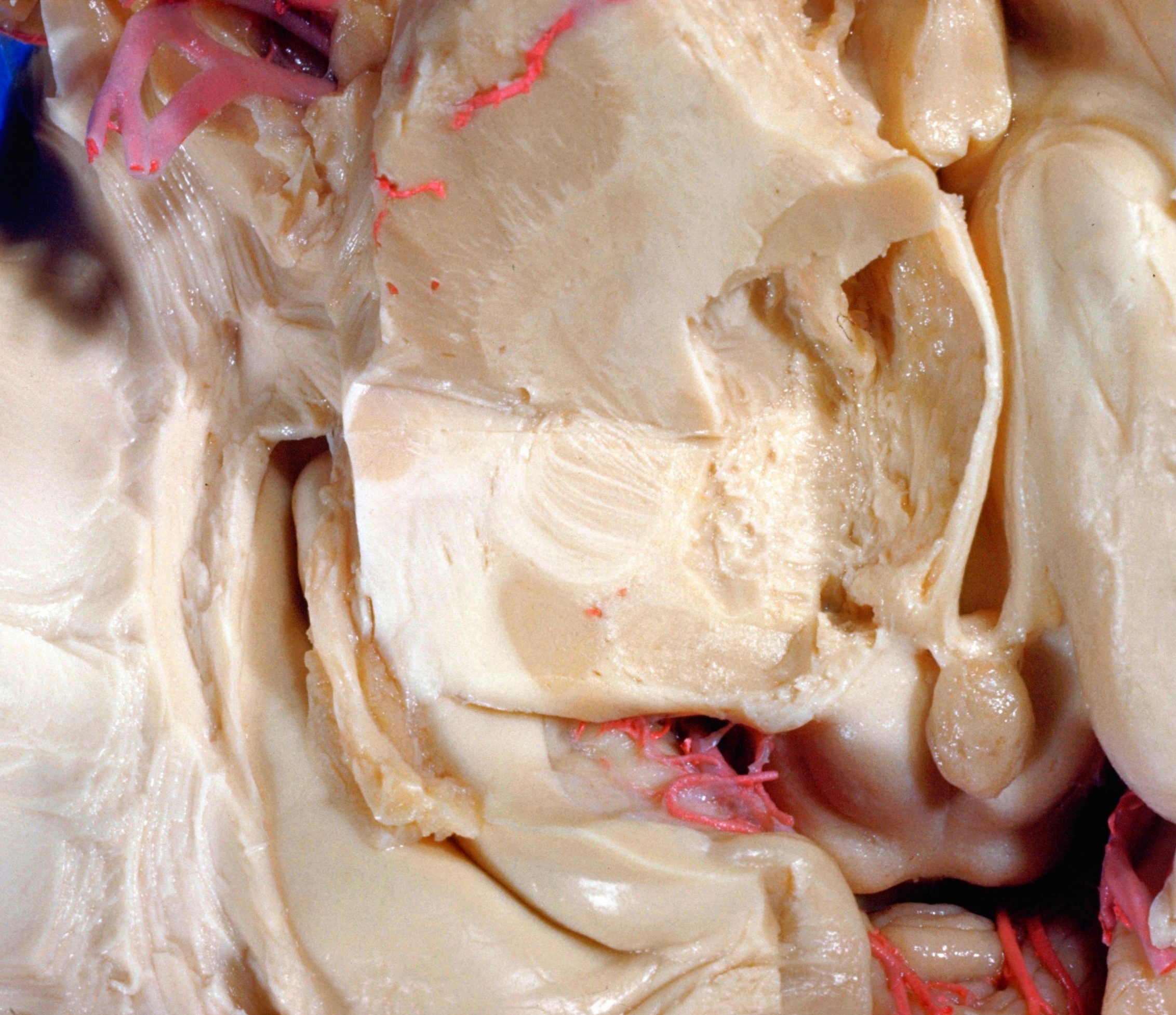
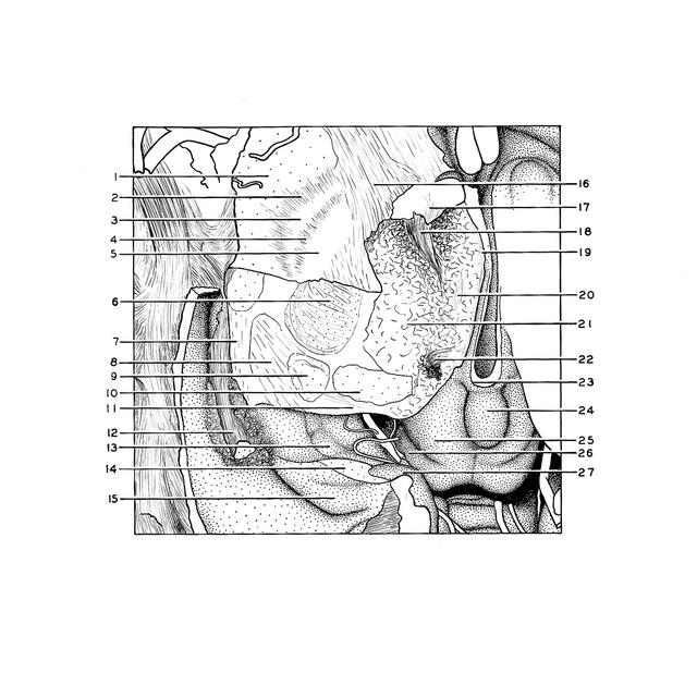
Close-up view of the preceding step in the dissection. The lateral geniculate body is sectioned horizontally, lamination within the nucleus being visible. The superior quadrigeminal brachium (11), connecting the optic tract to the superior colliculus, appears as an inconspicuous white band along the posterior margin of the cut section. The medial geniculate body is also cut across in a horizontal plane. The course of the inferior quadrigeminal brachium (26) towards this nucleus is partially hidden by its passage beneath the superior quadrigeminal brachium. The H fields in the subthalamic region (21) are not readily demonstrated by gross dissection although the fibrous nature of the area is apparent in the view. (The red nucleus lies immediately beneath this area.)
- Putamen
- External medullary lamina
- Globus pallidus (external division)
- Internal medullary Iamina
- Globus pallidus (internal division)
- Internal capsule (cut across)
- Fibers of stratum zonale of thalamus continuing toward temporal lobe
- Geniculocalcarine tract
- Lateral geniculate body
- Medial geniculate body
- Superior quadrigeminal brachium
- Stria terminalis
- Dentate fascia
- Fornix (ems) (cut across)
- Hippocampus
- Internal capsule (note many fibers passing from thalamic region into capsule at this point, comprising part of the frontal stalk of the thalarnus)
- Anterior nucleus of thalamus
- Mamillothalamic tract (Vicq d'Azyr)
- Stria medullaris thalami
- Periventricular fibers coursing towards mesencephalon
- H field of Forel (subthalamus)
- Fasciculus retroflexus
- Taenia thalami
- Pineal body
- Superior colliculus
- Inferior quadrigeminal brachium
- Limbic lobe (directly inferior to splenium of corpus callosum removed at an earlier phase of the dissection)