Exploration of the brain from its superior aspect
Anterior ventral nucleus of thalamus; H field of Forel; retrolenticular part of internal capsule
Stanford holds the copyright to the David L. Bassett anatomical images and has assigned
Creative Commons license Attribution-Share
Alike 4.0 International to all of the images.
For additional information regarding use and permissions,
please contact the Medical History Center.
Image #12-1
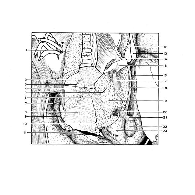
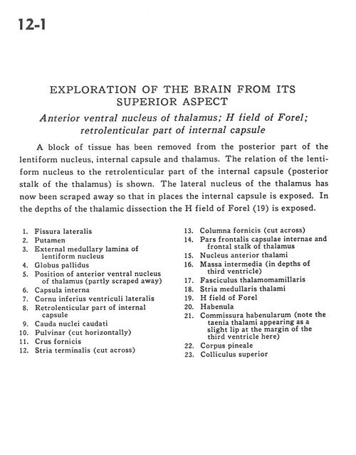
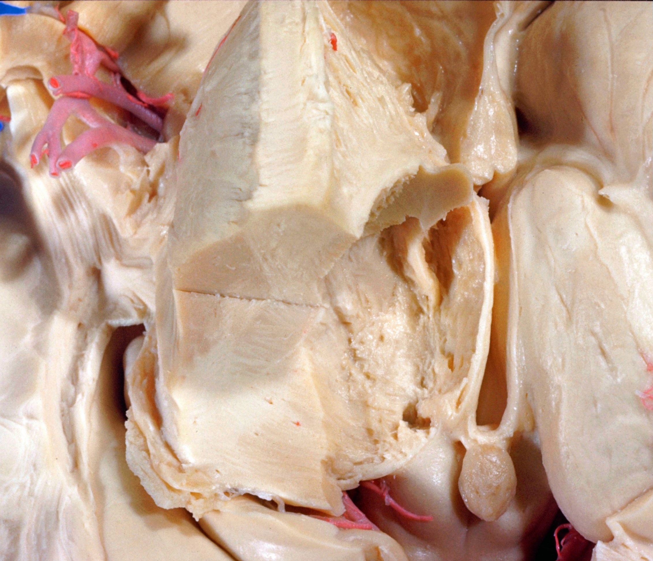
Exploration of the brain from its superior aspect
Anterior ventral nucleus of thalamus; H field of Forel; retrolenticular part of internal capsule
A block of tissue has been removed from the posterior part of the lentiform nucleus, internal capsule and thalamus. The relation of the lentiform nucleus to the retrolenticular part of the internal capsule (posterior stalk of the thalamus) is shown. The lateral nucleus of the thalamus has now been scraped away so that in places the internal capsule is exposed. In the depths of the thalamic dissection the H field of Forel (19) is exposed.
- Lateral fissure
- Putamen
- External medullary lamina of lentiform nucleus
- Globus pallidus
- Position of anterior ventral nucleus of thalamus (partly scraped away)
- Internal capsule
- Inferior horn of lateral ventricle
- Retrolenticular part of internal capsule
- Caudate nucleus (tail)
- Pulvinar (cut horizontally)
- Fornix (crus)
- Stria terminalis (cut across)
- Column of fornix (cut across)
- Frontal part internal capsule and frontal stalk of thalamus
- Anterior nucleus of thalamus
- Massa intermedia (in depths of third ventricle)
- Mamillothalamic tract
- Stria medullaris thalami
- H field of Forel
- Habenula
- Habenular commissure (note the taenia thalami appearing as a slight lip at the margin of the third ventricle here)
- Pineal body
- Superior colliculus