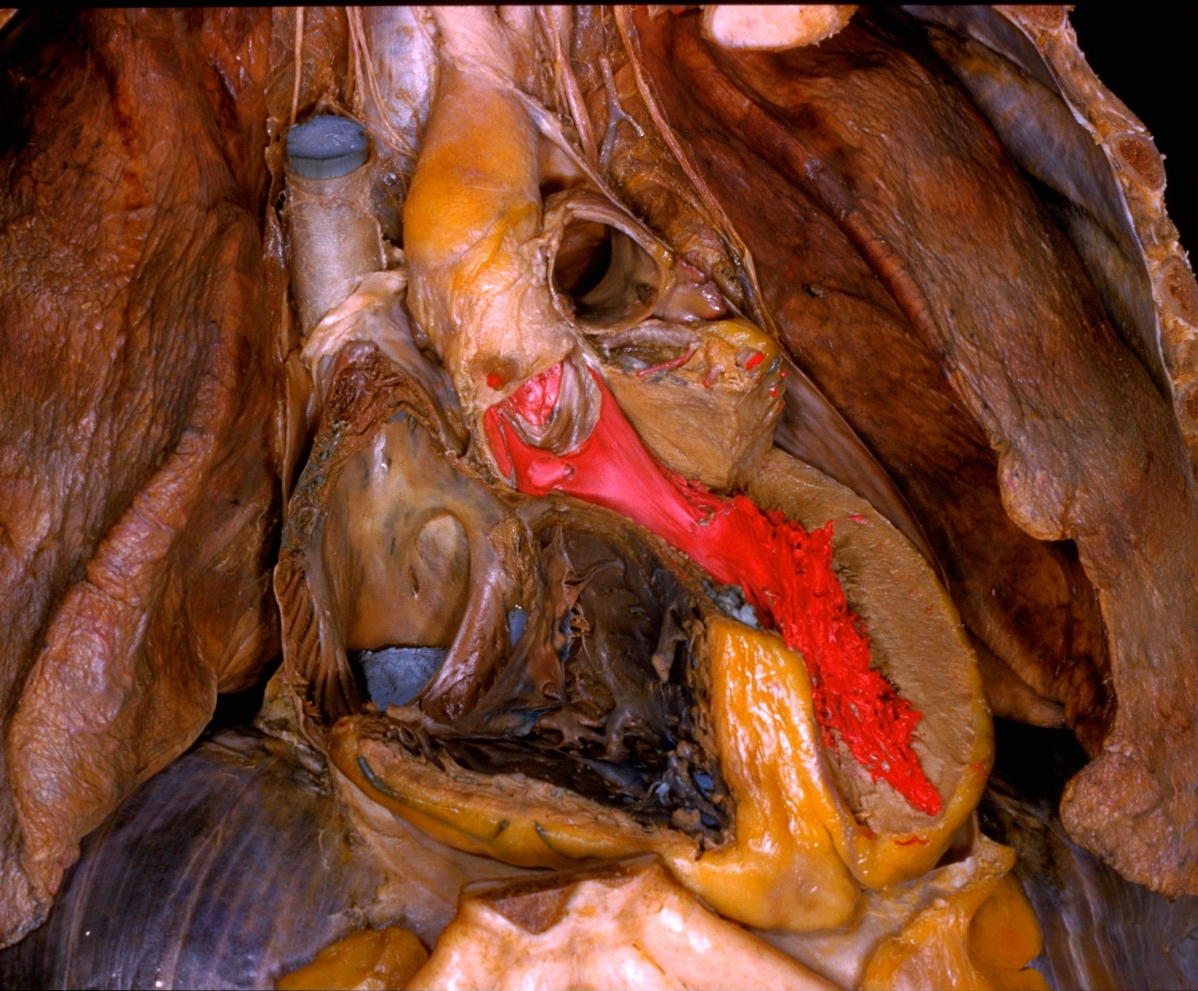
Dissection of pericardium and heart in situ
Conduction system of heart.
Stanford holds the copyright to the David L. Bassett anatomical images and has assigned
Creative Commons license Attribution-Share
Alike 4.0 International to all of the images.
For additional information regarding use and permissions,
please contact the Medical History Center.
Image #118-2
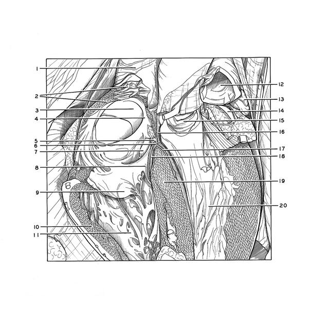
Dissection of pericardium and heart in situ
Conduction system of heart.
The camera angle has been adjusted to provide a cross-sectional view of the interventricular septum (19). This septum has been retracted slightly away from the left ventricle and has been cut in such a way that the course of the atrioventricular bundle (6) can be traced from its origin in the A-V node (5) to its division into right and left branches (17, 18) at the junction of the membranous and muscular parts of the interventricular septum. The parts of the conduction system have been colored white to provide better photographic contrast.
- Superior vena cava
- Cut edge of wall of right atrium (note sinusoidal spaces filled with latex and continuous with cavity of atrium)
- Fossa ovalis
- Border of fossa ovalis
- Atrioventricular node (colored white)
- Trunk of atrioventricular bundle (colored white)
- Coronary sinus
- Septal (medial) cusp of tricuspid valve
- Posterior cusp tricuspid valve
- Myocardium of right ventricle
- Right ventricle
- Pulmonary trunk
- Left auricle
- Left coronary artery
- Aortic valve (left semilunar cusp)
- Membranous part interventricular septum (partially resected to display A-V bundle)
- Left crus atrioventricular bundle (colored white)
- Right crus atrioventricular bundle (colored white)
- Muscular part interventricular septum
- Latex cast in cavity of left ventricle